. 2025 Mar 8;44(1):87.
doi: 10.1186/s13046-025-03348-0.

Low-coverage whole genome sequencing of cell-free DNA to predict and track immunotherapy response in advanced non-small cell lung cancer

Affiliations

Low-coverage whole genome sequencing of cell-free DNA to predict and track immunotherapy response in advanced non-small cell lung cancer

Florian Janke et al. J Exp Clin Cancer Res. .

Abstract

Background: Outcomes under anti-PD-(L)1 therapy have been variable in advanced non-small cell lung cancer (NSCLC) without reliable predictive biomarkers so far. Targeted next-generation sequencing (NGS) of circulating tumor DNA (ctDNA) has demonstrated potential clinical utility to support clinical decisions, but requires prior tumor genetic profiling for proper interpretation, and wide adoption remains limited due to high costs.

Methods: Tumor-agnostic low-coverage ctDNA whole genome sequencing (lcWGS) was used to longitudinally track genome-wide copy number variations (CNVs) and fragmentation features in advanced NSCLC patients (n = 118 samples from 49 patients) and healthy controls (n = 57). Tumor PD-L1 expression was available for comparison.

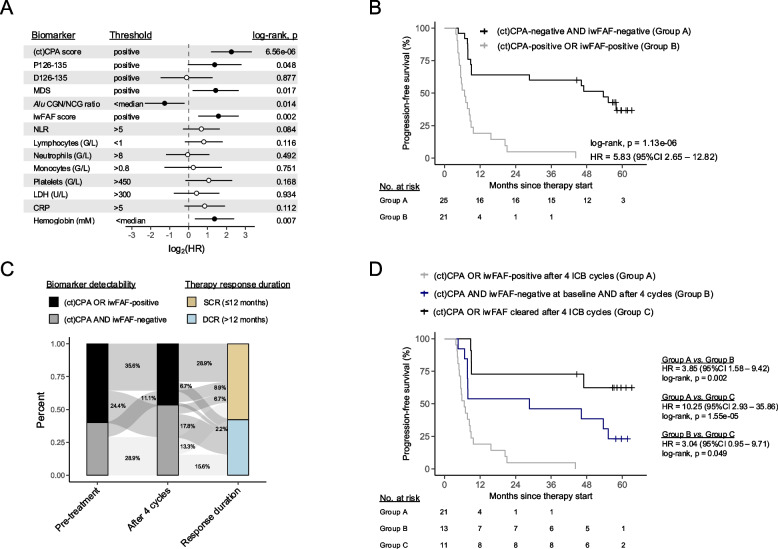
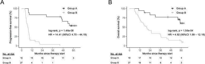
Findings: Fragmentation features and CNVs were complementary indicators, whose combination significantly increased ctDNA detection compared to single-marker assessments (+ 20.3% compared to CNV analysis alone). Baseline fragment length alterations, but not CNVs, were significantly associated with subsequent progression-free survival (PFS; hazard ratio [HR] = 4.10, p = 6.58e-05) and could improve PFS predictions based on tumor PD-L1 expression alone (HR = 2.70, p = 0.019). Residual CNVs or aberrant fragmentation of ctDNA under ongoing therapy could stratify patients according to the subsequent response duration (median 5.8 vs. 47.0 months, p = 1.13e-06). The integrative analysis of ctDNA fragment characteristics at baseline, tumor PD-L1 expression, and residual ctDNA under ongoing treatment constituted the strongest independent predictor of PFS (p = 6.25e-05) and overall survival (p = 1.3e-03) in multivariable analyses along with other clinicopathologic variables.

Interpretation: This study demonstrates the feasibility and potential clinical utility of lcWGS for the tumor-agnostic stratification and monitoring of advanced NSCLC under PD-(L)1 blockade based on CNV and fragmentomic profiling.

Keywords: CfDNA fragmentation; Copy number variations; Immunotherapy; Liquid biopsy; Low-coverage whole genome sequencing; Non-small cell lung cancer; Response prediction.


Conflict of interest statement

Declarations. Ethics approval and consent to participate: This study was approved by the ethics committee of the Heidelberg University (S-270/2001, S-296/2016, S-145/2018) and performed in accordance with the Declaration of Helsinki. Written informed consent was obtained from all participants prior to study inclusion. Consent for publication: Not applicable. Competing interests: The authors declare no competing interests.

Figures

Fig. 1
Fig. 1
A Copy number abnormality (CPA) scores in healthy donors compared to pre-treatment NSCLC patients. The number of samples surpassing the 95% specificity threshold and the total number of samples per group are given below the graph. B Fragment length distribution of CPA+ (n = 27), CPA pre-treatment NSCLC samples (n = 21), and the median size profile of all healthy donors (n = 57). 10-bp periodicity segments are indicated by the broken vertical lines. The correlation between the proportion of fragments between 126 and 135-bp (P126-135; shaded area) and CPA scores is highlighted on the right. C Comparison of fragmentation features (i.e., P126-135, absolute P126-135 deviation from the median of healthy donors [D126-135], motif diversity scores (MDS), Alu element methylation based on CGN/NCG end motif ratios, and information-weighted fraction of aberrant fragments (iwFAF) scores) between healthy donors, CPA+ and CPA pre-treatment NSCLC samples
Fig. 2
Fig. 2
A Oncoprint highlighting the detectability of circulating tumor DNA (ctDNA) based on cell-free copy number variations (CNVs; CPA and ctCPA score), cell-free DNA (cfDNA) fragment length (P126-135 and D126-135), fragment end motif diversity scores (MDS), fragment end-based DNA methylation inference (Alu CGN/NCG ratio), and fragment end position (iwFAF score). Colored tiles indicate detectable ctDNA at 95% specificity assessing the respective cfDNA biomarker. The absolute and relative number of ctDNA+ samples is given on the right side of the plot. B ctDNA detectability per sampling timepoint, illustrating the added value of joint CNV and cfDNA fragmentation analysis. Colors indicate the evaluation of CPA scores alone, the number of additional ctDNA+ samples when ctCPA scores were analyzed in addition to CPA scores, and when cfDNA fragmentation was assessed in addition to CPA and ctCPA scores. cfDNA fragmentation-positivity refers to the detectability of at least one of the 5 evaluated features. CPA, copy number abnormality; ctCPA, ctDNA-informed CPA; iwFAF, information-weighted fraction of aberrant fragments; PD-L1, programmed death-ligand 1; TPS, tumor proportion score
Fig. 3
Fig. 3
A Forest plot reporting the hazard ratios (HRs) of disease progression and their 95% confidence intervals for cfDNA biomarkers at baseline, clinically relevant PD-L1 tumor proportion scores (TPS), and different routine laboratory values. Statistically significant results are indicated by filled dots. Threshold to dichotomize patients are given for each biomarker. ‘Positive’ refers to detectable levels of the respective biomarker according to its 95% specificity threshold. Hemoglobin and creatinine concentrations are not illustrated due to group sizes < 4. Progression-free survival (PFS) according to the detectability (at 95% specificity) of D126-135 (B) and PD-L1 tumor proportion scores (TPS) ≥ 1% (C) at anti-PD-(L)1 therapy baseline. D Association between PFS and the combination of D126-135 detectability and/or PD-L1 TPS < 1% at therapy baseline. Groups were compared by two-sided log-rank tests and hazard ratios were calculated via univariate Cox proportional hazard models. CRP, C-reactive protein; D126-135, deviation of fragments between 126 and 135-bp from healthy donors; iwFAF, information-weighted fraction of aberrant fragments; LDH, lactate dehydrogenase; MDS, motif diversity score; NLR, neutrophil-to-lymphocyte ratio; P126-135, proportion of fragments between 126 and 135-bp; PD-L1, programmed death-ligand 1
Fig. 4
Fig. 4
A Forest plot of progression-free survival (PFS) univariate Cox regression for cfDNA biomarkers and routine laboratory values measured after 4 therapy cycles. Filled dots represent significant results. Patients were grouped based on the indicated thresholds. ‘Positive’ refers to biomarkers exceeding its respective 95% specificity threshold. Comparisons with group sizes < 4 are not included. B Association between PFS and combined evaluation of (ct)CPA and iwFAF scores detectability after therapy cycle 4. C Distribution of patients with and without residual ctDNA (evaluated via (ct)CPA and iwFAF score detecability) before and after 4 therapy cycles, showing the association of biomarkers status (i.e., detectable vs. undetectable) and duration of response. D Kaplan–Meier curve comparing PFS in patients with undetectable (ct)CPA and iwFAF scores at baseline and after 4 therapy cycles to patients that clear marker detectability and patients that remain biomarker positive at follow-up. Groups were compared by two-sided log-rank tests and hazard ratios (HR) were calculated via univariate Cox proportional hazard models. CNV, copy number variation; CRP, C-reactive protein; ctCPA, ctDNA-informed copy number abnormality; ctDNA, circulating tumor DNA; DCR, durable clinical response; iwFAF, information-weighted fraction of aberrant fragments; LDH, lactate dehydrogenase; MDS, motif diversity score; NLR, neutrophil-to-lymphocyte ratio; SCR, short clinical response
Fig. 5
Fig. 5
Comparison of progression-free (PFS; A) and overall survival (OS, B) between patient groups separated according to the following criteria: Group A; PD-L1 tumor proportion score (TPS) ≥ 1%, undetectable baseline D126-135 levels, as well as undetectable (ct)CPA and iwFAF scores after 4 therapy cycles. Group B; PD-L1 TPS < 1% or marker detectability for at least one of the evaluated cfDNA biomarkers. cfDNA biomarker detectability was assessed using the markers respective 95% specificity threshold. Groups were compared using two-sided log-rank tests, and hazard ratios (HR) were determined through univariate Cox proportional hazards models. cfDNA, cell-free DNA; ctCPA, ctDNA-informed copy number abnormality; D126-135, deviation of fragments between 126 and 135-bp from healthy donors; iwFAF, information-weighted fraction of aberrant fragments; PD-L1, programmed death-ligand 1

References

    1. Borghaei H, Paz-Ares L, Horn L, Spigel DR, Steins M, Ready NE, et al. Nivolumab versus Docetaxel in Advanced Nonsquamous Non-Small-Cell Lung Cancer. N Engl J Med. 2015;373(17):1627–39. - PMC - PubMed
    1. Reck M, Rodríguez-Abreu D, Robinson AG, Hui R, Csőszi T, Fülöp A, et al. Pembrolizumab versus Chemotherapy for PD-L1-Positive Non-Small-Cell Lung Cancer. N Engl J Med. 2016;375(19):1823–33. - PubMed
    1. Jenkins RW, Thummalapalli R, Carter J, Cañadas I, Barbie DA. Molecular and Genomic Determinants of Response to Immune Checkpoint Inhibition in Cancer. Annu Rev Med. 2018;69:333–47. - PubMed
    1. Rizvi H, Sanchez-Vega F, La K, Chatila W, Jonsson P, Halpenny D, et al. Molecular Determinants of Response to Anti-Programmed Cell Death (PD)-1 and Anti-Programmed Death-Ligand 1 (PD-L1) Blockade in Patients With Non-Small-Cell Lung Cancer Profiled With Targeted Next-Generation Sequencing. J Clin Oncol. 2018;36(7):633–41. - PMC - PubMed
    1. Rizvi NA, Hellmann MD, Snyder A, Kvistborg P, Makarov V, Havel JJ, et al. Cancer immunology. Mutational landscape determines sensitivity to PD-1 blockade in non-small cell lung cancer. Science. 2015;348(6230):124–8. - PMC - PubMed

MeSH terms