. 2025 Mar 8;74(4):143.
doi: 10.1007/s00262-025-03997-2.

Combination therapy with expanded natural killer cells and atezolizumab exerts potent antitumor immunity in small cell lung cancer

Affiliations

Combination therapy with expanded natural killer cells and atezolizumab exerts potent antitumor immunity in small cell lung cancer

Manh-Cuong Vo et al. Cancer Immunol Immunother. .

Abstract

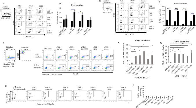
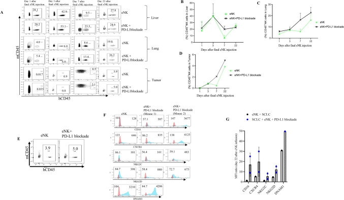
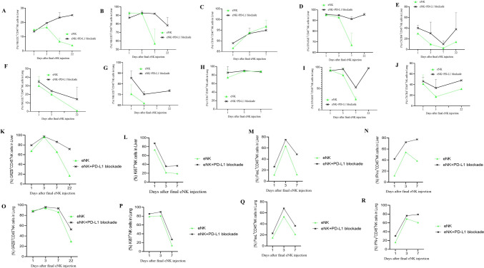
Despite an initial response to platinum-based chemotherapy, most patients with extensive stage of small cell lung cancer (SCLC) have a poor prognosis due to recurrence. Additionally, the benefit of immune checkpoint inhibitors is more modest than non-small cell lung cancer. Natural killer (NK) cells can directly eliminate cancer cells without prior sensitization; this is largely governed by inflammatory cytokines, which serve as killing signals to cancer cells. Here, we investigated whether the combination of NK cells plus atezolizumab, a fully humanized monoclonal antibody that specifically targets the protein programmed death-ligand 1 (PD-L1), has a synergistic effect against SCLC. NK cells were expanded and activated using irradiated K562 feeder cells in the presence of interleukin (IL)-2/IL-15/IL-21/41BB ligand for 14 days. Expanded and activated NK cells (eNK) were combined with atezolizumab and used to treat SCLC cells in both in vitro and in vivo studies. The results revealed increased PD-L1 expression in SCLC cells after the eNK challenge. eNK cells plus atezolizumab demonstrated increased cytotoxicity toward target SCLC cells, as evidenced by increased interferon-γ and tumor necrosis factor-α production, and higher levels of SCLC stem cell (CD44+CD90+) suppression. Combined treatment with eNK and atezolizumab more effectively inhibited SCLC tumor growth and significantly prolonged the survival of treated mice. Our findings revealed that combining eNK with atezolizumab strongly increased cytotoxicity, significantly inhibited SCLC tumor growth, and prolonged the survival of treated mice. These results provide a framework for developing a more advanced immunotherapeutic modality for future clinical trials for patients with SCLC.

Keywords: Anti-PD-L1; Atezolizumab; Combination therapy; NK cells; Small cell lung cancer.


Conflict of interest statement

Declarations. Conflicts of interest: The authors declare that they have no conflicts of interest.

Figures

Fig. 1
Fig. 1
Significant upregulation of PD-L1 molecules on the surface of SCLC and eNK cells after coculture. GFP-positive SCLC cell lines, including SBC-5, SHP77, DMS273, H69, and H209, were co-cultured with eNK cells to evaluate PD-L1 expression levels on both SCLC and eNK cells. PD-L1 expression on SCLC cells was assessed at 6 h of co-culture (A and B) and following overnight co-culture (C and D) using flow cytometry. Similarly, PD-L1 expression on eNK cells after co-culture with SCLC cell lines was measured at 6 h (E and F) and overnight (E and G) using flow cytometry. Differences between groups were analyzed using two-way ANOVA. (* P < 0.05, ** P < 0.012, *** P < 0.001). (H and I) PD-L1 expression on eNK cells after cultured in transwell with SCLC cells. Data are representative from three independent experiments (n = 3)
Fig. 2
Fig. 2
PD-1 is upregulated on the surface of SCLC and eNK cells after co-culture. GFP-positive SCLC cell lines, including SBC-5, SHP77, DMS273, H69, and H209, were co-cultured with eNK cells to evaluate the percentages of PD-1 expression on both SCLC cell lines and eNK cells. (A and B) PD-1 expression on SCLC cell lines was analyzed after 6-h and (C and D) overnight co-culture with eNK cells using flow cytometry. Similarly, PD-1 expression on eNK cells was assessed following co-culture with SCLC cell lines at (E and F) 6-h and (E and G) overnight time points, also measured by flow cytometry. Differences between groups were analyzed using two-way ANOVA. *P < 0.05, **P < 0.012, and ***P < 0.001. (H and I) PD-1 expression on eNK cells co-cultured with SCLC cell lines in transwell systems was analyzed. Data are representative from three independent experiments (n = 3)
Fig. 3
Fig. 3
eNK cells plus PD-L1 blockade greatly suppressed the stem cell population, as well as the neuroendocrine (NE) and non-neuroendocrine cell phenotypes (ML) coexisting in SCLC. (A and B) Quantification of stem cell-like markers (CD44 and CD90) expressed on small cell lung cancer (SCLC) cells, as assessed by flow cytometry. (C and D) Analysis of the co-expression of CD56 (NE) and CD44 (ML) markers in SCLC cell lines, evaluated using flow cytometry. Differences between groups were analyzed using two-way ANOVA. (*P < 0.05). Data are representative from three independent experiments (n = 3)
Fig. 4
Fig. 4
PD-L1 blockade robustly increases the cytotoxicity of eNK cells against SCLC cell lines. (A) The killing capacity of eNK cells against SCLC was investigated using an LDH-release cytotoxicity assay. (B) FACS plots and (C) Bar graphs illustrating the cytotoxic activity of eNK cells against CFSE-labelled SCLC cell lines. (D) FACS analysis and (E) Bar graph quantification of CD107a expression in eNK following coculture with target SCLC cell lines. (F) FACS analysis and (G) Bar graph showing the percentage of IFN-γ.+ eNK following coculture with target SCLC cell lines. Differences between groups were analyzed using two-way ANOVA. Data are representative from three independent experiments (n = 3)
Fig. 5
Fig. 5
eNK cells plus PD-L1 blockade exerts potent anticancer effects in the SCLC xenograft model. (A) Schematic summarizing the treatment of SCLC-bearing mice with eNK cells combined with PD-L1 blockade. (B) Representative images of injected SCLC tumors from mice (7 mice per group) treated with eNK cells plus PD-L1 blockade. (C) Growth rates of the tumor mass (****P < 0.0001), and (D) survival time of tumor-bearing mice (**P < 0.01). (E) Serum levels of pro-gastrin-releasing peptide (ProGRP) in the serum of vaccinated mice (**P < 0.012, ****P < 0.0001). Differences between groups were analyzed using two-way ANOVA. Data are representative from two independent experiments
Fig. 6
Fig. 6
PD-L1 blockade improves the in vivo effector function and persistence of eNK cells in SCLC-bearing mice. (A) Dot plots showing the in vivo distribution of CD45+ eNK cells in the liver, lung, and tumor of vaccinated mice. Bar graphs quantifying the distribution of circulating eNK cells in the liver (B), lung (C), and tumor (D), assessed via flow cytometry. On day 22 after final eNK cell infusion, mice were sacrificed, and single cell suspensions from the tumor were used to assess the effector function of tumor infiltrating eNK cells. (E) Representative FACS dot plot showing the in vivo distribution of hCD45+ eNK cells in the tumor of vaccinated mice. (F) FACs histograms provide the expression of activation markers on eNK cells in the tumor of vaccinated mice, including memory receptors such as NKG2C, activation receptors such as CD16 and NKG2D, migration receptors such as CXCR4, and costimulatory molecules such as DNAM-1. (G) The MFI ratios (MFI of samples/MFI of isotype controls) of each sample are shown as bar graphs. Data are derived from two representative mice (n = 2)
Fig. 7
Fig. 7
PD-L1 blockade improves in vivo cytotoxicity function of eNK cells in SCLC-bearing mice. Bar graphs showing the phenotypic characteristics of circulating eNK in the lung and liver, including memory receptors such as NKG2C (A and F), activation receptors such as NKG2D (B and G) and CD16 (C and H), costimulatory molecules such as DNAM-1 (D and I), and migration receptors such as CXCR4 (E and J). Data are derived from two representative mice (n = 2). Bar graphs quantifying the levels of cytotoxicity markers in of circulating eNK cells in the liver and lung of vaccinated mice, such as granzyme-B (K and O), proliferation markers such as Ki67 (L and P), apoptosis ligands such as FasL (M and G), and IFN-γ (N and R). Data are derived from single representative mice at each time point (n = 1)

References

    1. Lee JG, Kim HC, Choi CM (2021) Recent trends of lung cancer in Korea. Tuberc Respir Dis (Seoul) 84(2):89–95. 10.4046/trd.2020.0134 - PMC - PubMed
    1. Remon J, Aldea M, Besse B, Planchard D, Reck M, Giaccone G, Soria JC (2021) Small cell lung cancer: a slightly less orphan disease after immunotherapy. Ann Oncol 32(6):698–709. 10.1016/j.annonc.2021.02.025 - PubMed
    1. Charles MR, Elisabeth B, Corinne FF, Julien S (2021) Small-cell lung cancer. Nat Rev Dis Primers 7(1):3. 10.1038/s41572-020-00235-0 - PMC - PubMed
    1. Dingemans AMC, Früh M, Ardizzoni A, Besse B, Faivre-Finn C, Hendriks LE et al (2021) Small-cell lung cancer: ESMO Clinical Practice Guidelines for diagnosis, treatment and follow-up. Ann Oncol 32(7):839–853. 10.1016/j.annonc.2021.03.207 - PMC - PubMed
    1. van Weverwijk A, de Visser KE (2023) Mechanisms driving the immunoregulatory function of cancer cells. Nat Rev Cancer 23(4):193–215. 10.1038/s41568-022-00544-4 - PubMed

MeSH terms