. 2025 Mar 18;6(3):102019.
doi: 10.1016/j.xcrm.2025.102019. Epub 2025 Mar 7.

Engineered human myogenic cells in hydrogels generate innervated vascularized myofibers within dystrophic mouse muscle on long-term engraftment

Affiliations

Engineered human myogenic cells in hydrogels generate innervated vascularized myofibers within dystrophic mouse muscle on long-term engraftment

Anna Kowala et al. Cell Rep Med. .

Abstract

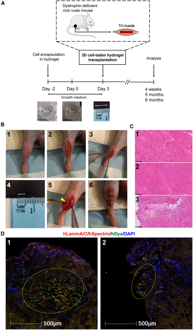
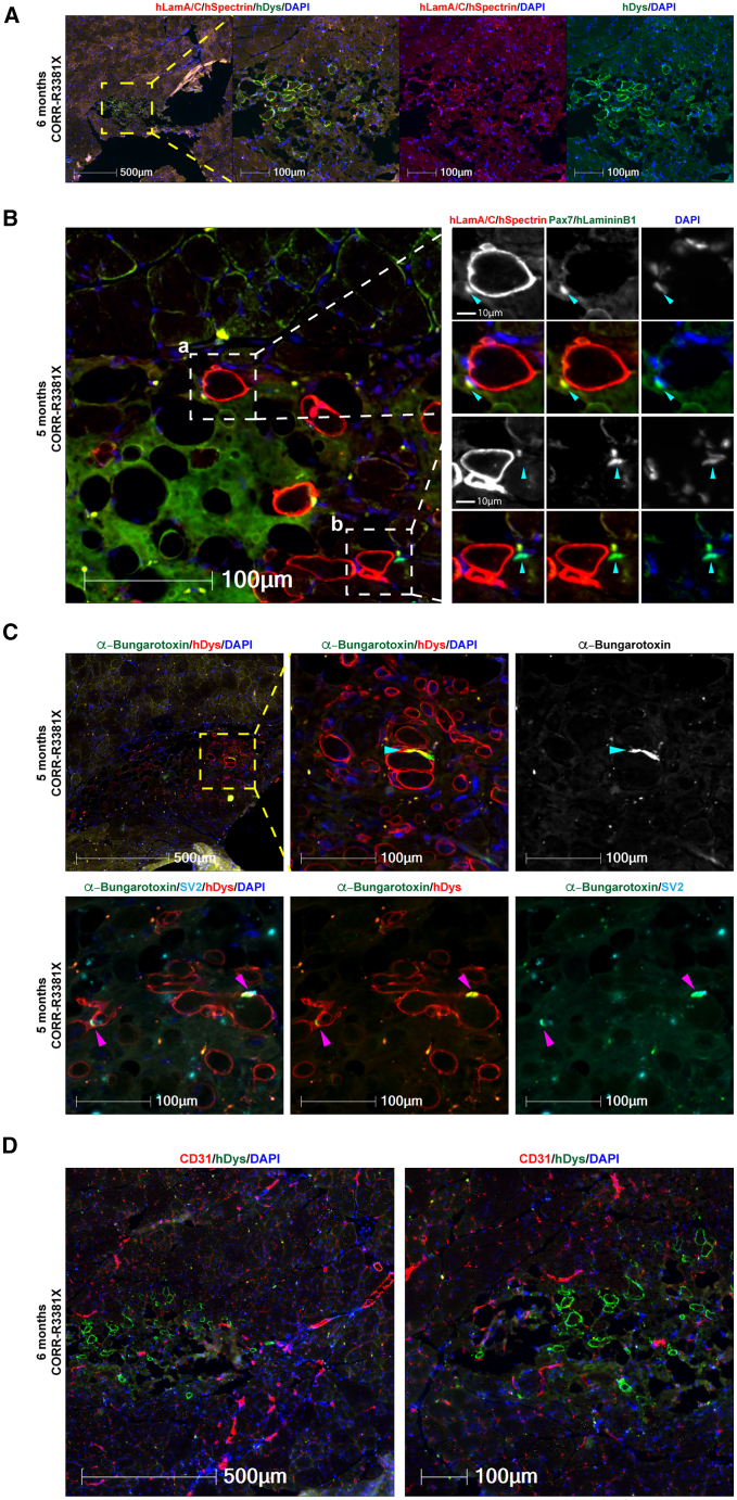
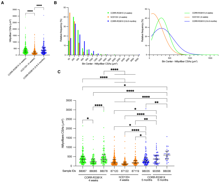
Transplantation of human myogenic progenitor cells (MPCs) is a promising therapeutic strategy for treating muscle-wasting diseases, e.g., Duchenne muscular dystrophy (DMD). To increase engraftment efficiency of donor stem cells, modulation of host muscles is required, significantly limiting their clinical translation. Here, we develop a clinically relevant transplantation strategy synergizing hydrogel-mediated delivery and engineered human MPCs generated from CRISPR-corrected DMD patient-derived pluripotent stem cells. We demonstrate that donor-derived human myofibers produce full-length dystrophin at 4 weeks and 5-6 months (long-term) after transplantation in the unmodulated muscles of the dystrophin-deficient mouse model of DMD. Remarkably, human myofibers are innervated by mouse motor neurons forming neuromuscular junctions and supported by vascularization after long-term engraftment in dystrophic mice. PAX7+ cells of human origin populate the satellite cell niche. There was no evidence of tumorigenesis in mice engrafted with hydrogel-encapsulated human MPCs. Our results provide a proof of concept in developing hydrogel-based cell therapy for muscle-wasting diseases.

Keywords: CRISPR; Duchenne muscular dystrophy; biomaterials; dystrophin; hydrogels; innervation; regenerative medicine; stem cells; vascularization; xenoengraftment.


Conflict of interest statement

Declaration of interests Y.-Y.L. was the Principal Investigator in a research project funded by Pfizer. O.P. is a co-founder and shareholder of Somite Therapeutics.

Figures

None
Graphical abstract
Figure 1
Figure 1
Signaling pathways required for muscle stem cell function are differentially enriched in DMD MPCs compared to CRISPR-corrected MPCs (A) Heatmap of all DE genes between undifferentiated DMD and CORR MPCs (day 0, cultured in growth medium). FDR < 0.05. 3 biological replicates for the DMD-R3381X and CORR-R3381X isogenic pair and 4 biological replicates for the DMD-K2957fs and CORR-K2957fs isogenic pair. See also Data S1. (B) Bubble plot of selected signaling pathways from gProfiler analysis. Bubbles sized by pathway size. See also Data S2. (C) Bubble plot of normalized enrichment scores (NESs) of selected gene sets negatively and positively enriched in DMD versus CORR MPCs from GSEA (adjusted p < 0.05). Bubbles sized by pathway size. See also Data S3. (D) Volcano plot shows DE genes (DMD versus CORR) with genes of interest annotated. DE genes with an FDR < 0.05 and a log fold change <−1.5 or >1.5 are labeled red, while genes only meeting the FDR threshold are labeled blue.
Figure 2
Figure 2
Experimental timeline and xenoengraftment of engineered human 3D cell-laden constructs in mdx nude mice (A) Following encapsulation of human PSC-derived MPCs in hydrogel in PDMS molds, 3D cell-laden constructs were cultured for 5 days in growth medium (either Promocell or Megacell) and then transplanted into TA muscles of mdx nude mice, followed by analysis at 4 weeks, 5 months, or 6 months after xenoengraftment. Scale bars, 10 mm. (B) Step-by-step transplantation procedure: (1) positioning of the hindlimb; (2) skin incision to reveal TA muscle; (3) TA muscle incision with a scalpel; (4) prior to transplantation, hydrogel with encapsulated cells was cut with 5 mm-diameter biopsy punch and removed from its PDMS mold; (5) placement of a 3D construct into TA muscle; and (6) closure of the muscle incision, with a 3D construct inside, and suture of the skin. (C) Representative H&E staining of transverse cryosections of TA muscles. Hematoxylin (purplish blue) stains cell nuclei, and eosin (pink) stains the extracellular matrix and cytoplasm. Panel 1, C57Bl/10 mouse (non-dystrophic control) with nuclei located at the periphery of myofibers; panel 2, non-transplanted mdx nude (dystrophic control). Central nuclei within the myofibers are characteristic of mdx nude mouse muscles. Panel 3, at 4 weeks after transplantation, the transplanted hydrogel construct (dashed yellow circle) is visible within mdx nude TA muscle. Scale bars, 100 μm. (D) Representative images of hydrogel-based engraftment of human myogenic cells in TA muscle of mdx nude mice at 4 weeks after transplantation. Engrafted cells and myofibers of human origin in the middle of mouse TA muscle (dashed yellow circles, panel 1) or at the edge of mouse TA muscle (dashed yellow circles, panel 2) are shown. Transverse 10 μm sections were stained with antibodies against human lamin A/C and human spectrin (both red) and human dystrophin (green). Nuclei were counterstained with DAPI (blue). Scale bars, 500 μm.
Figure 3
Figure 3
Engrafted cells and myofibers of human origin in mdx nude mice at 4 weeks after transplantation (A) Representative transverse cryosections of mdx nude mouse TA muscle transplanted with 3D constructs of CORR-R3381X MPCs in Promocell growth medium, CORR-K2957fs MPCs in Promocell growth medium, CORR-R3381X MPCs in Megacell growth medium, CORR-K2957fs MPCs in Megacell growth medium, and hCD133+ cells in Megacell growth medium. At 4 weeks after transplantation, transverse 10 μm sections were stained with antibodies against human lamin A/C and human spectrin (both red) and human dystrophin (green). Nuclei were counterstained with DAPI (blue). Scale bars, 100 μm. (B) Quantification of myofibers and undifferentiated cells of human origin in each experimental condition or independent replicate (Rep 1, 2, and 3). Schematics represent immunostaining patterns considered as myofibers of human origin (hMyofibers) or undifferentiated cells of human origin (hLamin A/C+ only). Values indicate minimum, maximum, median, and 25th and 75th percentiles. (C) Pie charts show percentages of engrafted human myogenic cells in mdx nude mice as undifferentiated cells and differentiated human myofibers. CORR-R3381X MPCs in Promocell growth medium (15 TA muscles), CORR-K2957fs MPCs in Promocell growth medium (12 TA muscles), and hCD133+ cells in Megacell growth medium (6 TA muscles). In Promocell medium: CORR-R3381X, 15 biological replicates. CORR-K2957fs, 12 biological replicates. In Megacell medium: CORR-R3381X, 6 biological replicates. CORR-K2957fs, 3 biological replicates; hCD133+, 6 biological replicates.
Figure 4
Figure 4
Distribution patterns of donor-derived human myofibers in mdx nude mice at 4 weeks after transplantation (A) Representative transverse cryosections of donor-derived human myofibers derived from cell-laden constructs of CORR-R3381X MPCs or CORR-K2957fs MPCs in Promocell growth medium and hCD133+ cells in Megacell growth medium. Human lamin A/C and human spectrin (both red) and human dystrophin (green). Nuclei were counterstained with DAPI (blue). Scale bars, 10–500 μm as indicated in each panel. (B) Pie charts show percentages of distribution patterns of donor-derived human myofibers in mdx nude mice. CORR-R3381X MPCs in Promocell growth medium (15 TA muscles), CORR-K2957fs MPCs in Promocell growth medium (12 TA muscles), and hCD133+ cells in Megacell growth medium (6 TA muscles).
Figure 5
Figure 5
Long-term engraftment of human myofibers and human PAX7+ cells in the satellite cell niche with innervation and vascularization in mdx nude mice (A) A representative transverse cryosection of mdx nude mouse TA muscle transplanted with a 3D construct of CORR-R3381X MPCs in Promocell growth medium (6 months after transplantation). Human lamin A/C and human spectrin (both red) and human dystrophin (green). Nuclei were labeled with DAPI (blue). (B) Detection of human (a) and mouse (b) PAX7+ cells (arrowheads) in the satellite cell compartment at 5 months after transplantation. (a) A PAX7+ cell (green) of human origin labeled by human laminA/C (red) and DAPI (blue) adjacent to a human myofiber (hSpectrin, red) beneath the basal lamina (hLaminin β1, green). (b) A PAX7+ cell (green) of mouse origin with DAPI (blue) adjacent to a human myofiber (hSpectrin, red) within the basal lamina (hLaminin β1, green). (C) Innervation of donor-derived human myofibers (hDystrophin, red) in mdx nude mice at 5 months after transplantation. The formation of NMJs (arrowheads) is demonstrated by co-localization of post-synaptic marker AChR (labeled by α-Bungarotoxin, green) and pre-synaptic marker SV2 (cyan). Nuclei were labeled with DAPI (blue). (D) Vascularization in the engrafted regions at 6 months after transplantation as demonstrated by detection of blood vessels (CD31, red) adjacent to human myofibers (hDystrophin, green). Nuclei were labeled with DAPI (blue). 6 biological replicates for long-term engraftment experiment. Scale bars, 10–500 μm as indicated in each panel.
Figure 6
Figure 6
CORR-R3381X MPC-derived human myofibers matured further upon long-term engraftment (A) Comparison of cross-section areas (CSAs) of donor-derived hMyofibers in mouse TA muscles from three experimental conditions, CORR-R3381X 4 weeks (391 CSAs from 3 samples), hCD133+ 4 weeks (518 CSAs from 3 samples), and CORR-R3381X 5 and 6 months (185 CSAs from 3 samples). Values indicate median (25th–75th percentile). One-way ANOVA and Tukey’s multiple comparisons test; ∗∗∗∗p < 0.0001. (B) Relative frequency distribution (%) of CSAs of hMyofibers derived from CORR-R3381X MPCs (4 weeks), hCD133+ cells (4 weeks), and CORR-R3381X MPCs (5 and 6 months), which were then fitted with non-linear regression curves to indicate the trends in each condition. (C) Comparisons of CSA measurements from individual TA muscle samples that ranked in top 3 engraftment efficiencies in each experimental condition. Values indicate median (25th–75th percentile). One-way ANOVA and Tukey’s multiple comparisons test; ∗p < 0.05; ∗∗p < 0.01; ∗∗∗∗p < 0.0001.

References

    1. Mauro A. SATELLITE CELL OF SKELETAL MUSCLE FIBERS. J. Biophys. Biochem. Cytol. 1961;9:493–495. doi: 10.1083/jcb.9.2.493. - DOI - PMC - PubMed
    1. Montarras D., Morgan J., Collins C., Relaix F., Zaffran S., Cumano A., Partridge T., Buckingham M. Direct isolation of satellite cells for skeletal muscle regeneration. Science. 2005;309:2064–2067. doi: 10.1126/science.1114758. - DOI - PubMed
    1. Yin H., Price F., Rudnicki M.A. Satellite Cells and the Muscle Stem Cell Niche. Physiol. Rev. 2013;93:23–67. doi: 10.1152/physrev.00043.2011. - DOI - PMC - PubMed
    1. Mercuri E., Bönnemann C.G., Muntoni F. Muscular dystrophies. Lancet. 2019;394:2025–2038. doi: 10.1016/S0140-6736(19)32910-1. - DOI - PubMed
    1. Boldrin L., Zammit P.S., Morgan J.E. Satellite cells from dystrophic muscle retain regenerative capacity. Stem Cell Res. 2015;14:20–29. doi: 10.1016/j.scr.2014.10.007. - DOI - PMC - PubMed

LinkOut - more resources