. 2025 Mar 8;8(1):395.
doi: 10.1038/s42003-025-07742-4.

Transcription factor ELF-1 protects against colitis by maintaining intestinal epithelium homeostasis

Affiliations

Transcription factor ELF-1 protects against colitis by maintaining intestinal epithelium homeostasis

Gege He et al. Commun Biol. .

Abstract

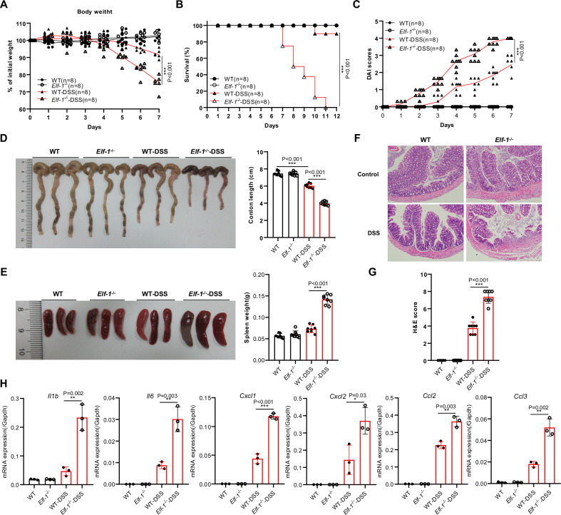
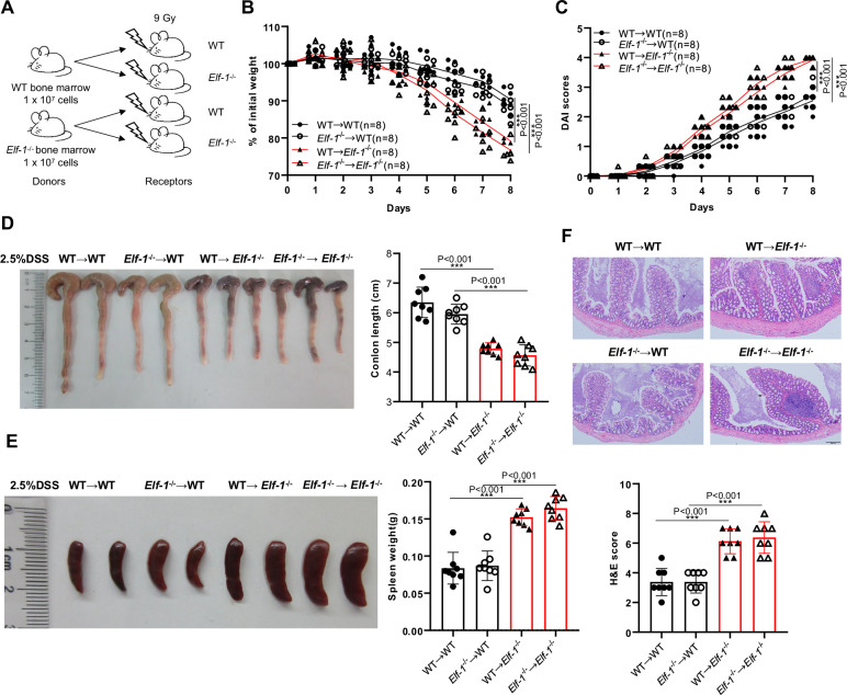
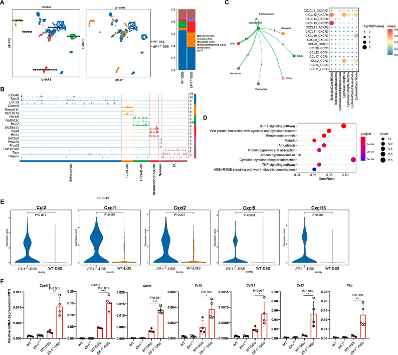
Inflammatory bowel disease (IBD) is a chronic, relapsing, and remitting disease characterized by chronic inflammation in the gastrointestinal tract. The exact etiology and pathogenesis of IBD remain elusive. Although ELF-1 has been known to be highly expressed in epithelial cells for past twenty years, little is known about its function in epithelial cells and epithelial-related IBD. Here, we demonstrated that ELF-1 deficiency in mouse lead to exacerbated DSS-induced colitis, marked by inflammation dominated by neutrophil infiltration and activation of IL-17 signaling pathways in various immune cells, including Th17, ILC3, γδT and NKT cells. Bone marrow transfer experiments confirmed ELF-1 deficiency in non-hematopoietic cells intrinsically worsened DSS-induced colitis. On one hand, ELF-1 deficiency enhanced the production of pro-inflammatory chemokines in colonic epithelial cells, leading to extensive infiltration of neutrophils and other immune cells into the colonic mucosal tissue. On the other hand, ELF-1 directly regulated the expression of the Rack1 gene in colonic epithelial tissue, which has been proved to play critical roles in maintaining intestinal homeostasis. Altogether, ELF-1 plays a protective role in colitis by maintaining intestinal epithelium homeostasis.


Conflict of interest statement

Competing interests: The authors declare no competing interests.

Figures

Fig. 1
Fig. 1. ELF-1 deficiency aggravated colitis.
A The body weight of WT and Elf-1−/− mice under normal condition and DSS treatment (n = 8). The survival rate (B) and DAI scores (C) of WT and Elf-1−/− mice in DSS-induced colitis (n = 8). Colon lengths (D) and spleen weight (E) of WT and Elf-1−/− mice in DSS-induced colitis (n = 8). H&E staining (F) and H&E scores (G) of colon tissues of WT and Elf-1−/− mice under normal conditions and DSS treatment (n = 8). H The mRNA levels of Il1b, Il6, Cxcl1, Cxcl2, Ccl2 and Ccl3 in colonic tissues of WT and Elf-1−/− mice in DSS-induced colitis (n = 3). Data are shown as mean ± SD, representing one of at least three independent experiments. *P < 0.05, **P < 0.01, ***P < 0.001.
Fig. 2
Fig. 2. ELF-1 deficiency causing an inflammation situation dominated by neutrophil infiltration in DSS colitis.
A UMAP plots and proportions of the identified cell types from the colonic tissues of DSS-treated WT and Elf-1−/− mice. B Dotplot of top 3 marker genes for the different cell subsets as indicated. C Percentages of CD11b+Ly6G+ neutrophils in colonic tissues of DSS-treated WT and Elf-1−/− mice (n = 3). GO (D) and KEGG (E) analyses of up-regulated DEGs in neutrophils of DSS-treated Elf-1−/− mice. F Expressions of representative genes of TNF and IL-17 signaling pathways in neutrophils of DSS-treated WT and Elf-1−/− mice. The intensity of expression is indicated as specified by the color legend. G UMAP plots and proportions of identified six subtypes in neutrophils of DSS-treated WT and Elf-1−/− mice. H Tracksplot of top marker genes for the subpopulations of neutrophil as indicated. I GO analysis of up-regulated DEGs in two identified subtypes of neutrophils (neutrophils5 and neutrophils6). J Differences in pathways scored by GSVA among different neutrophils subtypes.
Fig. 3
Fig. 3. IL-17 signaling pathway was markedly upregulated in DSS-treaeted Elf-1−/− mice.
A UMAP plots and proportions of the identified T cell types from the colonic tissues of DSS-treated WT and Elf-1−/− mice. B Heatmap of top marker genes for the subpopulations of T cells and ILCs group as indicated. C Percentages of CD4+IL-17+ Th17 cells and γδ T cells in colonic tissues of DSS-treated WT and Elf-1−/− mice (n = 3). KEGG (D) and GSEA (E) analyses of up-regulated DEGs in Th17, ILC3, NKT and γδT cells of DSS-treated Elf-1−/− mice. F Heatmap of representative genes of IL-17 signaling pathways in Th17, ILC3, NKT and γδT cells of DSS-treated WT and Elf-1−/− mice. The intensity of expression is indicated as specified by the color legend.
Fig. 4
Fig. 4. ELF-1 deficiency in non-hematopoietic cells intrinsically aggravates DSS induced colitis.
A The schematic diagram of construction of bone marrow chimera. The body weight (B) and DAI scores (C) of irradiated WT and Elf-1−/− mice transferred with WT or Elf-1−/− bone marrow in DSS-induced colitis (n = 8). Colon lengths (D) and spleen weight (E) of irradiated WT and Elf-1−/− mice transferred with WT or Elf-1−/− bone marrow in DSS-induced colitis (n = 8). F H&E staining and H&E scores of irradiated WT and Elf-1−/− mice transferred with WT or Elf-1−/− bone marrow in DSS-induced colitis (n = 8). Data are shown as mean ± SD, representing one of at least three independent experiments. *P < 0.05, **P < 0.01, ***P < 0.001.
Fig. 5
Fig. 5. ELF-1 deficiency enhanced expression of chemokines in colonic epithelial cells in DSS-induced colitis.
A UMAP plots and proportions of the identified subtypes in colonic epithelial cells from the colonic tissues of DSS-treated WT and Elf-1−/− mice. B Tracksplot of top 3 marker genes for the subpopulations of colonic epithelial cells as indicated. C Ligand‒receptor interaction between colonic epithelial cell and immune cell types. D KEGG analysis of up-regulated DEGs in colonic epithelial cells of DSS-treated Elf-1−/− mice. E Expressions of representative genes of cytokine-cytokine receptor interaction signaling pathways in colonic epithelial cells of DSS-treated WT and Elf-1−/− mice. F The mRNA levels of Cxcl13, Cxcl2, Cxcl1, Ccl2, Ccl11, Ccl3 and Il1b in colonic epithelial tissues of normal and DSS-treated WT and Elf-1−/− mice (n = 4). Data are shown as mean ± SD, representing one of at least three independent experiments. *P < 0.05, **P < 0.01, ***P < 0.001.
Fig. 6
Fig. 6. ELF-1 directly regulates Rack1 expression in DSS colitis.
A Distribution of ELF-1 binding sites from ChIP-Seq analysis of colonic tissues of WT mice. B KEGG pathway enrichment analysis of ELF-1 binding genes (binding peaks located in gene promoter regions). C Venn diagram showing overlapping gene between ELF-1 binding genes with down regulated DEGs in colonic epithelial cells of DSS-treated Elf-1−/− mice. D ChIP-seq peak for ELF-1 binding site in Rack1 gene. E Top 2 predicted ELF-1 binding motifs in Rack1 gene promoter with the most significant P values. F Rack1 expressions in colonic epithelial cells of DSS-treated WT and Elf-1−/− mice. G Rack1 expressions in colonic epithelial tissues of normal and DSS-treated WT and Elf-1−/− mice (n = 4). Data are shown as mean ± SD, representing one of at least three independent experiments. **P < 0.01.

References

    1. de Souza, H. S. P., Fiocchi, C. & Iliopoulos, D. The IBD interactome: an integrated view of aetiology, pathogenesis and therapy. Nat. Rev. Gastroenterol. Hepatol.14, 739–749 (2017). - PubMed
    1. Flynn, S. & Eisenstein, S. Inflammatory Bowel Disease Presentation and Diagnosis. Surg. Clin. North Am.99, 1051–1062 (2019). - PubMed
    1. de Souza, H. S. & Fiocchi, C. Immunopathogenesis of IBD: current state of the art. Nat. Rev. Gastroenterol. Hepatol.13, 13–27 (2016). - PubMed
    1. Mayer, L. Evolving paradigms in the pathogenesis of IBD. J. Gastroenterol.45, 9–16 (2010). - PubMed
    1. Parikh, K. et al. Colonic epithelial cell diversity in health and inflammatory bowel disease. Nature567, 49–4 (2019). - PubMed

LinkOut - more resources