. 2025 May;39(5):1146-1156.
doi: 10.1038/s41375-025-02551-4. Epub 2025 Mar 8.

CD38 in the pathobiology of cutaneous T-cell lymphoma and the potential for combination therapeutic intervention

Affiliations

CD38 in the pathobiology of cutaneous T-cell lymphoma and the potential for combination therapeutic intervention

Colleen Isabelle et al. Leukemia. 2025 May.

Abstract

Cutaneous T-Cell Lymphoma (CTCL) is a non-Hodgkin's lymphoma involving malignant skin-homing T-cells, characterized by variable severity and limited treatment options. Our study shows that patient samples and derived cell lines express CD38 on CTCL cells, and αCD38 antibodies effectively target CD38 in a mouse model. In vivo αCD38 antibody treatment led to the loss of CD38 expression in residual tumor cells, highlighting the need for innovative strategies to improve CTCL outcomes despite the CD38 loss in residual tumor cells. To investigate the role of CD38 in CTCL pathology, we used CRISPR-Cas9 to create CD38-deficient (CD38KO) CTCL cells. These CD38KO cells showed higher expression of oncogenes B-catenin, TCF7, and BCL6, along with reduced migration. Elevated NAD+ levels in CD38KO cells increased cellular respiration after CD38 inhibition in CD38WT cells. In vivo, CD38KO cell transplants led to more aggressive tumors, likely due to elevated β-catenin, Bcl6, and Tcf-1 signaling. Prior research in multiple myeloma showed αCD38 antibody efficacy relies on CD38 expression. We discovered that panobinostat, a histone deacetylase inhibitor, increased surface CD38 expression in CTCL cells dose-dependently. Combining panobinostat with αCD38 antibody in a CTCL mouse model significantly improved survival compared to the antibody alone, underscoring CD38's therapeutic potential in CTCL. CD38 is expressed in CTCL cells and can be targeted with αCD38 antibody. αCD38 antibody treatment leads to a significant reduction in CTCL cells, while residual cells lose CD38 expression. Knocking out CD38 from CTCL cells leads to increases in intracellular NAD+ and increased cellular respiration. Additionally, CD38KO cells have increased protein levels of β-catenin, Tcf1 (encoded by TCF7), and Bcl6. CD38KO CTCL cells grow more aggressively in vivo than CD38WT CTCL cells. Treating CTCL cells with panobinostat increases CD38 expression. A dual combination treatment of panobinostat and αCD38 antibody in a mouse model of CTCL improved survival outcomes compared to αCD38 antibody treatment alone. (Figure made with Biorender.com).


Conflict of interest statement

Competing interests: The authors declare no competing interests. Ethics approval and consent to participate: All experiments were performed in accordance with relevant guidelines and regulations. All animal experiments were conducted with Institutional Animal Care and Use Committee (ID 02234) approval. Studies including human subjects or samples were approved by an Institutional Review Board (IRB 17D.083) and conducted in accordance with the Declaration of Helsinki with informed consent obtained from all subjects.

Figures
None
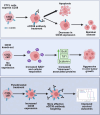
CD38 is expressed in CTCL cells and can be targeted with αCD38 antibody. αCD38 antibody treatment leads to a significant reduction in CTCL cells, while residual cells lose CD38 expression. Knocking out CD38 from CTCL cells leads to increases in intracellular NAD+ and increased cellular respiration. Additionally, CD38KO cells have increased protein levels of β-catenin, Tcf1 (encoded by TCF7), and Bcl6. CD38KO CTCL cells grow more aggressively in vivo than CD38WT CTCL cells. Treating CTCL cells with panobinostat increases CD38 expression. A dual combination treatment of panobinostat and αCD38 antibody in a mouse model of CTCL improved survival outcomes compared to αCD38 antibody treatment alone. (Figure made with Biorender.com).
Fig. 1
Fig. 1. CD38 expression in CTCL cells.
A CD38 expression in CTCL was assessed using data from Nielsen et al. 2021 (GSE143382), comparing relative CD38 expression in skin biopsy samples from CTCL patients (N = 70) to healthy donors (N = 12) (log fold change 4.8; p < 0.0001 by Mann–Whitney test). B Single-cell RNA sequencing analysis from previously published datasets (GSE128531, GSM5280111, GSE165623) [–14] compared CD38 cell expression in cells from healthy human skin (N = 4) to CTCL patient skin (N = 7). C CD38 expression in CTCL cell lines H9, HH, Hut78, and Hut102 was analyzed by flow cytometry. D Formalin-fixed, paraffin-embedded (FFPE) skin biopsies from CTCL patients (N = 6) and healthy donor skin (N = 1) were stained for CD38 and imaged at original magnification x4 and x20 using a Cytation5 imager and Gen5 software.
Fig. 2
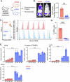
Fig. 2. αCD38 antibody treatment reduces CD38 expression and increases stemness genes in CTCL cells.
A Schematic representation of the experimental design for in vivo testing. CD38+ luciferase expressing CTCL cells (H9 line) were engrafted intravenously in immunodeficient NRG mice which were randomly assigned to treatment conditions with either αCD38 antibody (daratumumab; 100 mg/kg subcutaneously weekly for four weeks) or IgG isotype control (0.8 mg/kg subcutaneously once a week for four weeks). Tumor burden was measured over time using IVIS imaging and quantified using Living Image software. B Representative images of mice treated with isotype or αCD38 antibody and quantification of tumor burden total flux (photons/second) at 19 days post-engraftment. αCD38 antibody treatment resulted in an average total flux = 1.4e7 photons/sec (N = 4), while IgG average total flux = 9.0e7 photons/sec (N = 3); p = 0.0002. C Flow cytometry analysis of tumor cells isolated from the bone marrow of mice, performed 28 days post-engraftment after three weeks of treatment with either IgG isotype control (N = 5) or αCD38 (N = 7). Lymphocytes were gated for human CD45+ cells and analyzed for human CD38 signal. D Quantification of the percentage of CD45+ tumor cells expressing CD38 based on the data from panel (C) (p < 0.0001 by unpaired t test). E qPCR analysis of the relative expression of CD38, B-catenin (CTNNB1), TCF7, and BCL6 genes in CTCL tumor cells isolated from the bone marrow of mice that were treated with either isotype or αCD38 antibody (p < 0.0001 in all conditions by 2way ANOVA).
Fig. 3
Fig. 3. Comparison of growth, migration, and stemness protein expression in H9 CD38WT and CD38KO CTCL cell lines.
A Schematic demonstrating development process of CD38KO H9 CTCL cell line using CRISPR-Cas9, including flow cytometry showing post-sorting purity and CD38- status of CD38KO line (figure made with Biorender). B CD38KO CTCL cells showed no growth differences compared to CD38WT (p = 0.22 by linear regression). C Migration assay comparing CD38WT and CD38KO CTCL cells with and without CXCL12 chemoattractant (p < 0.01 by unpaired t test). D Western blot of β-catenin, Bcl6, and Tcf7 protein levels with β-actin loading control comparing between H9 CD38WT and H9 CD38KO luciferase CTCL cell lines and healthy donor CD4 + T-cells (HD CD4) imaged via chemiluminescence with a FluorChem Imager and images processed with AlphaView software.
Fig. 4
Fig. 4. Enhanced metabolic activity in CD38KO CTCL cell line and CD38WT cells following CD38 inhibitor treatment.
A Comparative assessment of total intracellular NAD+ and NADH levels between CD38WT and CD38KO CTCL cells using relative luminescence units (RLU) revealed significantly higher levels in CD38KO cells (p < 0.0001 by unpaired t test). B Separate measurements of NAD+ vs NADH levels in CD38WT and CD38KO CTCL cells by RLU indicated significantly elevated NAD+ levels in CD38KO cells (p < 0.0001 by unpaired t test). C Analysis of the NAD + /NADH ratio displayed a notable increase in CD38KO cells (p = 0.0006 by unpaired t test). D Seahorse XF Cell Mito Stress Test assay compared oxygen consumption rate (OCR) between the CD38WT and CD38KO CTCL cell lines (normalized to pmol/min/µg protein). E Quantification of the average baseline normalized OCR as well as maximal OCR from the seahorse assay in panel D indicated a significant increase in both baseline and maximal OCR in CD38KO cells compared to CD38WT cells (baseline OCR showed a 2.26-fold increase, p < 0.0001; maximal OCR exhibited a 1.83-fold increase, p < 0.0001 as determined by the Mann–Whitney test). F Seahorse XF Cell Mito Stress Test assay compared OCR of CD38WT cells treated with CD38 inhibitor compound 78c (0.5 µM overnight) or a DMSO control. G Quantification of baseline and maximal normalized OCR between CD38 inhibitor-treated cells and DMSO control cells demonstrated a significant increase in both baseline and maximal OCR in the CD38 inhibitor-treated cells (baseline OCR 1.38-fold increase, p < 0.0001; maximal OCR exhibited a 1.19 fold increase, p = 0.021 as determined by Mann–Whitney test).
Fig. 5
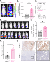
Fig. 5. Enhanced in vivo growth of CD38KO CTCL cells compared to CD38WT cells.
A Intravenously engrafted H9 CD38WT and H9 CD38KO CTCL cell lines in immunodeficient NOD Rag−/−γc−/− (NRG) mice were monitored over time using an In Vivo Imaging System (IVIS; Perkins-Elmer) and representative images from day 18 post-engraftment are shown. B Quantification of the tumor cell luminescent intensity signal as measured by total flux (photons/second) in the CD38WT and CD38KO intravenous cohorts (CD38WT = 2.2e8 photons/sec average total flux, N = 7; CD38KO = 1e9 photons/sec, N = 5; p = 0.003 by Mann–Whitney test). C Flow cytometry analysis of human CD45+ tumor cells harvested the bone marrow of NRG mice post IV-engraftment and expansion of H9 CD38WT and H9 CD38KO luciferase CTCL cell lines. D IVIS images of tumor signal at 19 days post-subcutaneous flank engraftment of H9 CD38WT and H9 CD38KO luciferase CTCL cell lines in NRG mice. E Tumor cell burden as measured by total flux of subcutaneous CD38WT and CD38KO cells (CD38WT = 1.01e8 photons/sec average total flux, N = 7; CD38KO = 6.01e9 photons/sec, N = 8; p < 0.0001 by unpaired t test). F Flank tumors were dissected and photographed on a grossing board. Image J was used to measure tumor size in mm2 (p = 0.01 by Mann–Whitney test). G Dissected tumors were formalin fixed and paraffin embedded before being stained for human CD38 and imaged at original magnification x4 and x20 on a Cytation5 imager with Gen5 software.
Fig. 6
Fig. 6. Panobinostat upregulates CD38 expression on H9 CTCL cells and enhances survival in a mouse model of CTCL in combination with αCD38 antibody immunotherapy.
A CTCL cells were treated with one of several histone deacetylase inhibitor agents followed by staining for CD38 and subsequent analysis via flow cytometry using a BD LSRFortessa (figure made with Biorender). B The mean fluorescent intensity of CD38 expression on CTCL cells treated for 72 h with either 1 μM vorinostat, 1 nM romidepsin, 25 nM of panobinostat, or DMSO control (panobinostat vs DMSO p < 0.001 by unpaired t test). C Representative histograms displaying CD38 expression at 72 h for increasing doses of panobinostat (5 nM, 10 nM, and 25 nM) compared to an isotype control. D Median fluorescence intensity (MFI) of CD38 expression on H9 cells treated with increasing doses of panobinostat for across multiple time points (24 h, 48 h, and 72 h) (max 85% increase in CD38 expression with 25 nM panobinostat vs. DMSO at 72 h; p < 0.0001 by 2way ANOVA). E Experimental design and treatment regimen for the in vivo combination αCD38 antibody and panobinostat in mice engrafted H9 luciferase CTCL cells. The animals were divided into four age and sex-matched experimental groups (N = 4 for all groups): Vehicle and IgG; Vehicle and αCD38 antibody; Panobinostat and IgG; and Panobinostat and αCD38 antibody. Panobinostat (20 mg/kg) and vehicle (2% DMSO, 48% PEG300, 2% Tween80, and 48% ddH2O) were administered via IP injection twice a week, while αCD38 antibody (daratumumab 100 mg/kg) and IgG isotype control (0.8 mg/kg) were administered subcutaneously once a week for three weeks. Mice were monitored and survival was tracked (figure made with Biorender). F Survival curve depicting the outcomes of the four experimental groups in the αCD38 antibody and panobinostat combination study (vehicle/IgG median survival 23 days; vehicle/ αCD38 antibody median survival 32 days; Panobinostat/IgG median survival 27.5 days; combination median survival 39 days; p = 0.01 by log-rank test).

References

    1. Agar NS, Wedgeworth E, Crichton S, Mitchell TJ, Cox M, Ferreira S, et al. Survival outcomes and prognostic factors in mycosis fungoides/Sézary syndrome: validation of the revised International Society for Cutaneous Lymphomas/European Organisation for Research and Treatment of Cancer staging proposal. J Clin Oncol. 2010;28:4730–9. - PubMed
    1. Duarte RF, Boumendil A, Onida F, Gabriel I, Arranz R, Arcese W, et al. Long-term outcome of allogeneic hematopoietic cell transplantation for patients with mycosis fungoides and Sézary syndrome: a European society for blood and marrow transplantation lymphoma working party extended analysis. J Clin Oncol. 2014;32:3347–8. - PubMed
    1. Isabelle C, Johnson WT, McConnell K, Vogel A, Brammer JE, Boles A, et al. Preclinical evaluation of anti-CD38 therapy in mature T-cell neoplasms. Blood Adv. 2023;7:3637–41. - PMC - PubMed
    1. Hogan KA, Chini CCS, Chini EN. The multi-faceted ecto-enzyme CD38: roles in immunomodulation, cancer, aging, and metabolic diseases. Front Immunol. 2019;10:1187. - PMC - PubMed
    1. Calabretta, E. & Carlo-Stella, C. The many facets of CD38 in lymphoma: from tumor-microenvironment cell interactions to acquired resistance to immunotherapy. Cells. 2020;9:802. - PMC - PubMed

MeSH terms