

Feasibility of shear wave elastography (2D -SWE) to evaluate cristalline lens in healthy dogs
- PMID: 40057718
- PMCID: PMC11889749
- DOI: 10.1186/s12917-025-04605-2
Feasibility of shear wave elastography (2D -SWE) to evaluate cristalline lens in healthy dogs
Abstract
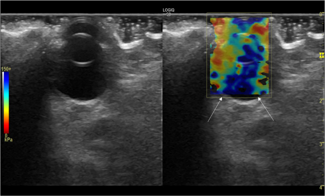
Background: 2D shear wave elastography (2D SWE) is an emerging technique in veterinary medicine able to assess tissue stiffness in a non-invasive way. Nowadays no report is yet available about its application in assessing the mechanical properties of canine lenses.
Objectives: This study aimed to evaluate the repeatability and reproducibility of 2D SWE in assessing normal lens elasticity in healthy and ageing dogs.
Methods: Trans-corneal lens 2D SWE was performed under physical restraint on 33 dogs by two operators who collected triplicate kPa and m/s measures, with the aim to assess reproducibility and reliability of the technique, followed by the evaluation of eventual difference of stiffness in different ages (G1 < 1.5 years, G2 1.5 years-7 years and G3 > 7 years). The project has been approved by the CEISA Ethical Committee (Prot. N. 12/2019 361 CEISA). Written informed consent was obtained by all the owners.
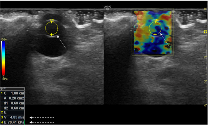
Results: Mean elasticity values were respectively 4.78 ± 1.48 m/s and 74.9 ± 43.7 kPa for the left eye and 4.45 ± 0.98 m/s and 75.9 ± 43.6 kPa for the right eye. Despite a slight difference observed in the measurements obtained in m/s between the two operators, the intra-observer assessment was excellent in the overall population of dogs for both values in KPa and m/s, as well as the inter-observer one (ICC > 0.75). All the sCV% computed evidence a low measurement dispersion (< 12%). Mean lens stiffness for G1 was 3.1 ± 0.5 m/s and 28.9 ± 9.3 kPa, for G2 4.61 ± 0.62 m/s and 65 ± 18.4 kPa and for G3 6.46 ± 0.36 m/s and 126 ± 14.5 kPa; a significant difference P (< 0.001) was detected between all the groups.
Conclusions: It can be concluded that 2D SWE is a rapid and non-invasive US-based technique able to assess lens mechanical properties in companion animals since it is characterized by high reliability and reproducibility, providing also information regarding lens stiffness in aging dogs.
Keywords: 2D shear wave elastography; Dog; Lens; US.
© 2025. The Author(s).
Conflict of interest statement
Declarations. Ethics approval and consent to participate: The project has been approved by the CEISA Ethical Committee (Committee on Animal Research and Ethics of the Universities of Chieti-Pescara, Teramo, 360 L’Aquila and of the Experimental Zooprophylactic Institute of Abruzzo-Molise, Prot. N. 12/2019 361 CEISA). Written informed owner’s consent was obtained before starting the procedure. Consent for publication: Not applicable. Competing interests: The authors declare no competing interests.
Figures

References
-
- Shiina T, Nightingale KR, Palmeri ML, Hall TJ, Bamber JC, Barr RG, et al. WFUMB guidelines and recommendations for clinical use of ultrasound elastography: part 1: basic principles and terminology. Ultrasound Med Biol. 2015;41(5):1126–47. - PubMed
-
- Bamber J, Cosgrove D, Dietrich CF, Fromageau J, Bojunga J, Calliada F, et al. EFSUMB guidelines and recommendations on the clinical use of ultrasound elastography. Part 1: basic principles and technology. Ultraschall Med. 2013;34(2):169–84. - PubMed
-
- Cintra CA, Feliciano MaR, Santos VJC, Maronezi MC, Cruz IK, Gasser B, et al. Applicability of ARFI elastography in the evaluation of canine prostatic alterations detected by b-mode and doppler ultrasonography. Arquivo Brasileiro De Med Veterinária E Zootecnia. 2020;72(6):2135–40.
-
- Cosgrove D, Piscaglia F, Bamber J, Bojunga J, Correas JM, Gilja OH, et al. EFSUMB guidelines and recommendations on the clinical use of ultrasound elastography. Part 2: clinical applications. Ultraschall Med. 2013;34(3):238–53. - PubMed
MeSH terms
LinkOut - more resources
Full Text Sources
