. 2025 Mar 8;24(1):72.
doi: 10.1186/s12943-025-02235-8.

Small extracellular vesicles and particles (sEVPs) derived from tumor-free pre-metastatic organs promote breast cancer metastasis and support organotropism

Affiliations

Small extracellular vesicles and particles (sEVPs) derived from tumor-free pre-metastatic organs promote breast cancer metastasis and support organotropism

Tasneem Cheytan et al. Mol Cancer. .

Abstract

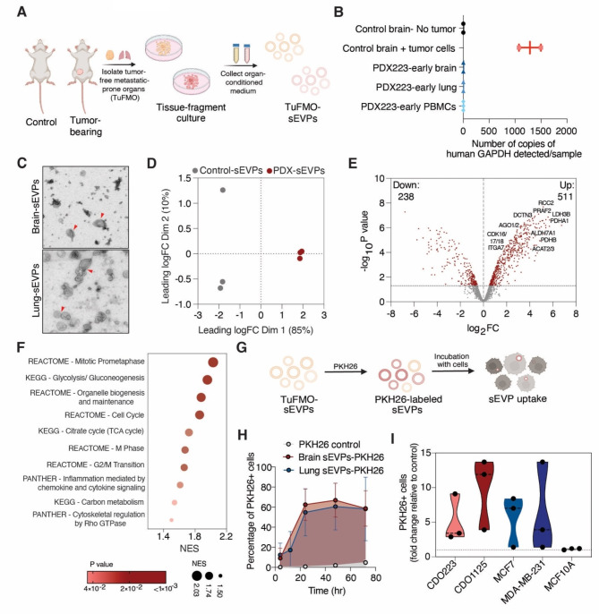
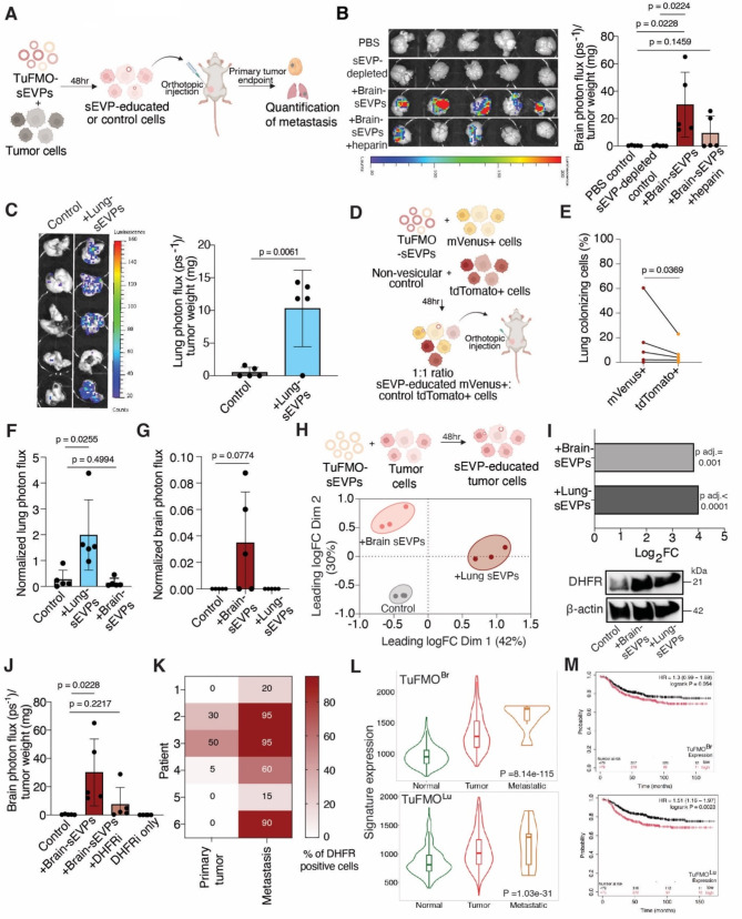
Metastatic breast cancer remains largely incurable, partly due to our incomplete understanding of its intricate underlying mechanisms. Notably, intercellular communication mediated by small extracellular vesicles and particles (sEVPs) has emerged as a key feature of metastasis. While tumor-derived sEVPs have been extensively studied and are known to be pro-metastatic, the role of sEVPs from metastasis-prone normal tissue sites remains primarily undefined. Here, we characterized and studied the function of sEVPs secreted from tumor-free pre-metastatic organs (TuFMO-sEVPs) such as the brain and lungs in both immunocompetent and patient-derived xenograft models. TuFMO-sEVPs from the brain of mammary tumor-bearing mice were found to have a distinct protein content as compared to brain-sEVPs from tumor-free mice, suggesting that the primary tumor can systemically influence the cargo of TuFMO-sEVPs. Importantly, mice orthotopically injected with breast cancer cells which had been educated with either brain or lung TuFMO-sEVPs prior to transplantation showed significantly increased metastasis to the respective organ. We further demonstrated that TuFMO-sEVPs induced the expression of the enzyme dihydrofolate reductase (DHFR) upon uptake by breast cancer cells, leading to their enhanced metastatic capacity. Organ-specific signatures generated from TuFMO-sEVP educated tumor cells were found to be increased in metastatic samples from breast cancer patients as compared to the primary tumor or normal tissue samples and these signatures also significantly correlated with poorer patient outcome. Collectively, our data reveals a novel facet of the metastatic cascade, implicating a role for TuFMO-sEVPs in directing metastasis and providing a potential therapeutic strategy for targeting this process.

Keywords: Brain metastasis; Breast cancer; Dihydrofolate reductase-mediated metastasis; Global proteomics analysis; Lung metastasis; Small extracellular vesicles and particles (sEVPs).


Conflict of interest statement

Declarations. Ethics approval and consent to participate: Animal experiments were performed in compliance with the German legal regulations and were approved by the governmental review board of the state of Baden-Württemberg in Germany, operated by the local Animal Welfare Office (Regierungspräsidium Karlsruhe) under the license number G-169/22. The use of patient-derived material for the establishment of PDX and CDOs used in this study was previously reported by our group (12) and the study was approved by the ethical committee of the University of Heidelberg (case number S-408/2013). Consent for publication: Not applicable. Competing interests: The authors declare no competing interests.

Figures

Fig. 1
Fig. 1
Tumor-free breast cancer pre-metastatic organs secrete sEVPs (TuFMO-sEVPs) which can be taken up by tumor cells. (A) Experimental scheme for the isolation of TuFMO-sEVPs from breast cancer pre-metastatic organs of patient-derived xenograft (PDX) and age-matched non-tumor-bearing control. (B) Digital droplet PCR analysis to detect human GAPDH in the intact brain, lungs or peripheral blood mononuclear cell (PBMC) fraction, n = 2–3 per condition. (C) Representative transmission electron microscopy (TEM) images of brain and lung TuFMO-sEVPs, indicated by the red arrow. (D) Principal component analysis (PCA) of the protein content of brain sEVPs from control mice vs. PDX based on global mass spectrometric analysis, n = 3 mice per group. (E) Volcano plot of differentially enriched proteins in brain TuFMO-sEVPs from PDX as compared to control mice (p < 0.05 labeled in red). (F) Gene set enrichment analysis (GSEA) of proteins significantly enriched in brain TuFMO-sEVPs from PDX as compared to control mice, ranked by normalized enrichment score (NES). (G) Experimental scheme to measure TuFMO-sEVP uptake by tumor cells through PKH26 labeling. (H) Uptake of 10 μg brain- and lung-derived TuFMO-sEVPs by MCF7 cells as measured by the percentage of PKH26 + cells over time, n = 3. (I) Uptake of 10 μg brain TuFMO-sEVPs by different breast cancer and breast epithelial cell lines and CDOs after 3 h, n = 3. Data information: Scale-bars: 250 nm (TEM). Data is represented as mean ± SD, all replicates shown are biological replicates
Fig. 2
Fig. 2
Brain and lung TuFMO-sEVPs promote brain and lung metastasis respectively. (A) Experimental scheme to determine the effect of TuFMO-sEVPs on metastasis. (B) Representative brain bioluminescence images (left) and brain metastatic burden (right) in mice orthotopically injected with CDO223 cells pre-educated with brain TuFMO-sEVPs with/without 10 μg/ml heparin, PBS or sEVP-depleted secretome, n = 5 mice per group. (C) Representative lung bioluminescence images (left) and lung metastatic burden (right) in mice orthotopically injected with CDO223 cells pre-educated with lung TuFMO-sEVPs or PBS (control), n = 5 mice per group. (D) Experimental scheme and (E) Percentage (%) of lung colonizing cells in mice orthotopically injected with a 1:1 ratio of lung sEVP-educated mVenus+ MDA-MB-231 cells: control tdTomato+ MDA-MB-231 cells, n = 5. (F) Lung and (G) Brain metastatic burden in BALB/c mice orthotopically injected with 4T1 cells pre-educated with TuFMO-sEVPs or PBS (control), n = 5 mice per group. (H) Experimental scheme (top) and PCA (bottom) of the proteomic profile of patient-derived CDO223 cells educated with TuFMO-sEVPs based on global mass spectrometric analysis, n = 3 per condition. (I) Log fold change showing the expression of DHFR from mass spectrometric-based proteomics analysis of CDO223 cells (top) or immunoblots of 4T1 cells (bottom) after education with brain or lung TuFMO-sEVPs. β-actin is the loading control. (J) Brain metastatic burden in mice orthotopically injected with CDO223 cells pre-educated with brain TuFMO-sEVPs with/without 10 μM pyrimethamine (DHFR inhibitor), treated with 10 μM pyrimethamine only or PBS (control), n = 5 mice per group. (K) Percentage of DHFR-positive tumor cells in paired breast cancer patient primary tumor and metastatic samples, n = 6. (L) Expression of the signature derived from CDO223 cells educated with brain (top) or lung (bottom) TuFMO-sEVPs in TNMplot breast invasive carcinoma dataset comparing metastatic vs. primary tumor vs. normal samples, p-value from Kruskal-Wallis test. (M) Kaplan Meier plots of the distant metastasis-free survival of breast cancer patients with low (black line) or high (red line) brain (TuFMOBr) (top) or lung (TuFMOLu) (bottom) signature using data from the Kaplan-Meier Plotter, n is indicated in each plot. Data information: 10 μg/ml sEVPs was used. Data is represented as mean ± SD, all replicates shown are biological replicates. Statistical significance was evaluated using two-tailed t-test (B, C, F, G, J) and two-tailed ratio paired t-test (E)

References

    1. Park M, Kim D, Ko S, Kim A, Mo K, Yoon H. Breast cancer metastasis: mechanisms and therapeutic implications. Int J Mol Sci. MDPI; 2022;23. - PMC - PubMed
    1. Wortzel I, Dror S, Kenific CM, Lyden D. Exosome-mediated metastasis: communication from a distance. Developmental Cell. Cell; 2019;49:347–60. - PubMed
    1. Peinado H, Alečković M, Lavotshkin S, Matei I, Costa-Silva B, Moreno-Bueno G, et al. Melanoma exosomes educate bone marrow progenitor cells toward a pro-metastatic phenotype through MET. Nat Med. 2012;18(6):883–91. - PMC - PubMed
    1. Blavier L, Nakata R, Neviani P, Sharma K, Shimada H, Benedicto A et al. The capture of extracellular vesicles endogenously released by xenotransplanted tumours induces an inflammatory reaction in the premetastatic niche. J Extracell Vesicles. 2023;12(5). - PMC - PubMed
    1. Kalluri R, LeBleu VS. The biology, function, and biomedical applications of exosomes. Science. American Association for the Advancement of Science. 2020;367. - PMC - PubMed

LinkOut - more resources