. 2025 Mar 8;26(1):92.
doi: 10.1186/s12931-025-03167-z.

Neutrophils are involved in the development and outcomes of plastic bronchitis associated with Mycoplasma pneumoniae pneumonia

Affiliations

Neutrophils are involved in the development and outcomes of plastic bronchitis associated with Mycoplasma pneumoniae pneumonia

Xia Huang et al. Respir Res. .

Abstract

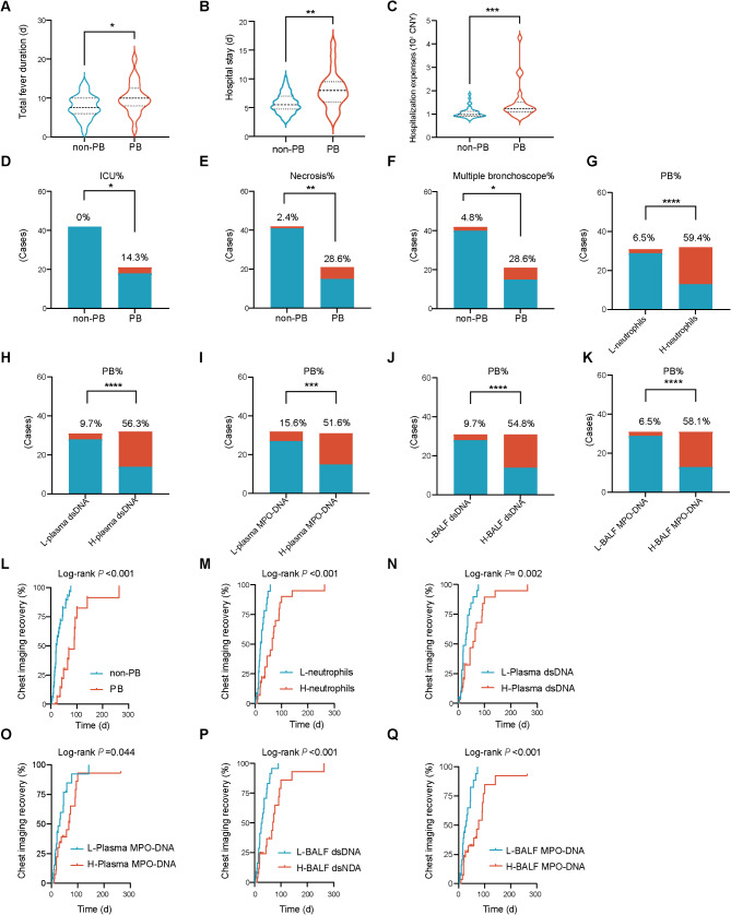
Background: Previous research has demonstrated a notable increase in neutrophil counts among pediatric patients with plastic bronchitis (PB) associated with Mycoplasma pneumoniae pneumonia (MPP). However, the role of neutrophils in MPP-associated PB remains largely elusive.

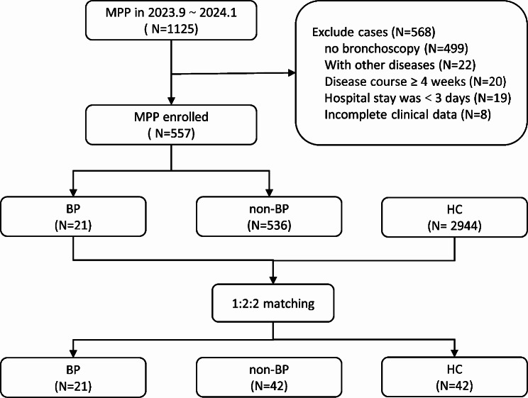
Methods: This is a nested case-control study that enrolled patients diagnosed with MPP who underwent bronchoscopy in our department during the MPP pandemic from September 2023 to January 2024. We conducted an analysis of clinical characteristics, blood samples, bronchoalveolar lavage fluid (BALF), and cast specimens, correlating these factors with the development and outcomes of PB.

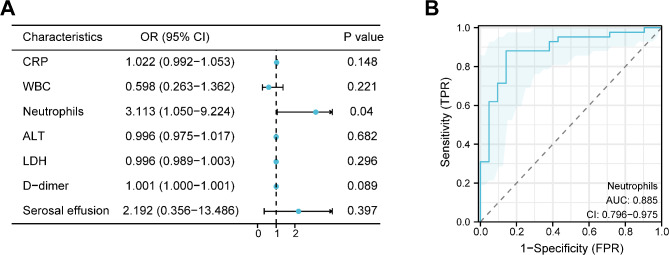
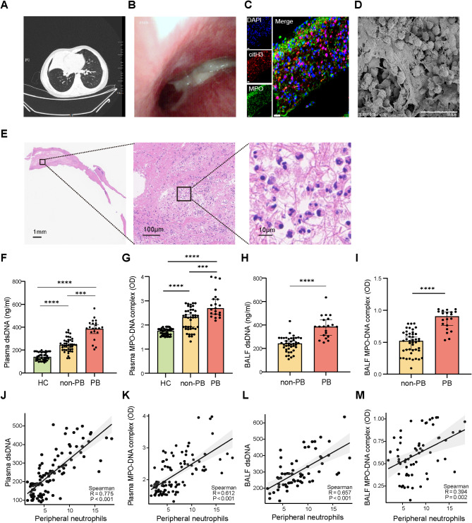
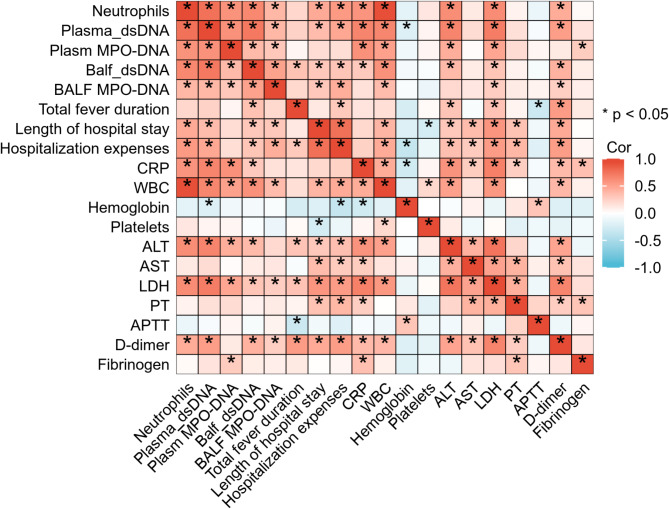
Results: Among the 557 patients with MPP included in the study, 21 (3.8%) developed PB. The peripheral neutrophil count was identified as an independent risk factor for PB (OR = 3.113 [95%CI 1.050-9.224], P = 0.04) and exhibited strong predictive value for the condition (AUC = 0.885 [95%CI 0.796-0.975], P < 0.001). Notably, there was a marked presence of neutrophil infiltration and neutrophil extracellular traps (NETs) formation in the blood, BALF, and cast samples from patients with PB. Furthermore, the levels of neutrophils and NETs correlated significantly with clinical outcomes.

Conclusion: A high level of neutrophils poses a risk for PB and demonstrates strong predictive value for its diagnosis. Neutrophils and NETs are closely linked to the clinical outcomes of PB in patients with MPP.

Keywords: Mycoplasma pneumoniae; Neutrophil extracellular traps; Neutrophils; Outcomes; Plastic bronchitis.


Conflict of interest statement

Declarations. Ethics approval: This study was approved by the research ethics committee of our institution (approval number: 202308005-1) and complied with the Declaration of Helsinki. Informed consent: The parents of all participating children provided written informed consent before inclusion in the study. Consent for consent for publication: Not applicable. Competing interests: The authors declare no competing interests.

Figures

Fig. 1
Fig. 1
Flowchart of the study. HC: heathy control; MPP: Mycoplasma pneumoniae pneumonia, PB: Plastic bronchitis
Fig. 2
Fig. 2
Risk assessment and predictive value of neutrophils for PB in MPP. (A) The forest plot derived from the multivariate logistic regression analysis illustrates that neutrophil counts independently predict the risk of PB development in the context of MPP. (B) The ROC curve analysis demonstrated that neutrophil counts possess a significant predictive value in the diagnosis of PB. ALT: alanine aminotransferase; AUC: area under the curve; CRP, C-reactive protein; CI: 95% confidence interval; LDH: lactate dehydrogenase; MPP: Mycoplasma pneumoniae pneumonia; OR: Odds Ratio, PB: Plastic bronchitis; ROC: receiver operating characteristic; WBC: white blood cell
Fig. 3
Fig. 3
Massive infiltration of neutrophils and formation of NETs in plasma, BALF, and bronchial casts were observed in MPP associated PB. (A) Chest computed tomography prior to bronchoscopy reveals consolidation and atelectasis in a single lobe of the lung. (B) Bronchoscopy revealed mucosal hyperemia and/or edema, as well as the presence of gelatinous, tree-like casts causing obstruction in the airway. (C) Representative immunofluorescence microscopy images depict the staining for MPO in green, citH3 in red, and DNA in blue within bronchial casts. The co-localization of DNA with both MPO and citH3 is evident, confirming the presence of NETs. n = 10. Scale bar = 20 μm. (D) A representative scanning electron microscopy image reveals the presence NET-like structures in close proximity to neutrophils. Scale bar = 30 μm. (E) Representative images of hematoxylin-eosin staining of the bronchial casts reveal chromatolysis and the presence of cell-free neutrophils, which are indicative of the formation of NETs. n = 10. Scale bar = 100 mm (left),100 μm (middle) and 10 μm (right). (F-G) Levels of dsDNA and MPO-DNA complex, biomarkers for the presence of NETs, were quantified in the plasma across three distinct study cohorts: patients diagnosed with PB (PB, n = 21), patients without PB (non-PB, n = 42), and healthy controls (HC, n = 42). (H-I) Levels of NETs were quantified in the BALF across two distinct study cohorts: patients diagnosed with PB (PB, n = 21), patients without PB (non-PB, n = 42). (J-M) Correlation curves were constructed to delineate the relationship between NETs levels in plasma and the counts of peripheral neutrophils, as well as between NETs levels in BALF and peripheral neutrophil counts Statistical analysis: (F-I) The graphical representation illustrates the median NETs levels, with each bar accompanied by its corresponding 95% CI. Statistical significance among the groups is indicated by asterisks, using the following convention: *P < 0.05, **P < 0.01, ***P < 0.005, ****P < 0.001. (H-I) The Spearman rank correlation test was utilized to evaluate the strength and direction of the association between plasma NETs concentrations and peripheral neutrophil counts. BALF: bronchoalveolar lavage fluid. citH3: citrullinated histone 3; CI: confidence interval; dsDNA: double-stranded DNA; MPO: myeloperoxidase; NETs: neutrophil extracellular traps; PB: plastic bronchitis
Fig. 4
Fig. 4
Analysis of the correlation between peripheral neutrophils, NETs and various clinical parameters. Statistical analysis: The Spearman correlation test was employed to measure the strength and direction of the association between two variables. In the graphical representation, red coloration is utilized to denote a positive correlation, whereas blue coloration signifies a negative correlation. * P < 0.05. ALT: alanine aminotransferase; AST: aspartate aminotransferase; APTT: activated partial thrombin time; BALF: bronchoalveolar lavage fluid, CRP, C-reactive protein; dsDNA: double-stranded DNA; LDH: lactate dehydrogenase; MPO: myeloperoxidase; PT: prothrombin time; WBC: white blood cell
Fig. 5
Fig. 5
Clinical outcomes of the patients with PB associated with MPP. (A-C) Violin plots illustrate the comparative analysis of the total fever duration, hospital stay, and hospitalization expenses between the PB and non-PB groups. (D-F) Cumulative bar graphs depict the comparative analysis of the proportions of ICU admissions, necrosis, and the requirement for multiple bronchoscopies between the PB and non-PB groups. (G-I) Cumulative bar graphs illustrate the comparisons of PB proportion between Low-neutrophils and High-neutrophils groups compare the proportions of PB across different groups, specifically between the Low-neutrophils and High-neutrophils groups (G), the Low-plasma dsDNA and High-plasma dsDNA groups (H), the Low-plasma MPO-DNA and High-plasma MPO-DNA groups (I), the Low- BALF dsDNA and High-BALF dsDNA groups (J), and the Low- BALF MPO-DNA and High-BALF MPO-DNA groups (K). (L-Q) Kaplan-Meier survival curves evaluate the time to chest imaging recovery, stratified by the presence of PB (L), neutrophil counts (M), plasma dsDNA (N), plasma MPO-DNA (O), BALF dsDNA (P) and BALF MPO-DNA (Q) Statistical analysis: Quantitative data with skewness were presented as the median with interquartile range (IQR: 25th–75th percentiles) and were subjected to the Wilcoxon-Mann-Whitney rank-sum test to assess statistical differences (A-C). Categorical data are expressed as frequencies and were evaluated using the chi-square test or Fisher’s exact test(D-K). *P < 0.05, **P < 0.01, ***P < 0.005, ****P < 0.001. The log-rank test was utilized to calculate the P values for comparing the survival distributions across these groups (L-Q). BALF: bronchoalveolar lavage fluid; H: high; ICU: intensive care unit; L: low; MPO: myeloperoxidase; NETs: neutrophil extracellular traps; PB: Plastic bronchitis

Similar articles

References

    1. Meyer Sauteur PM, Beeton ML. Mycoplasma pneumoniae: delayed re-emergence after COVID-19 pandemic restrictions. Lancet Microbe 2023. - PubMed
    1. Gong C, Huang F, Suo L, Guan X, Kang L, Xie H, Hu G, Yang P, Wang Q. Increase of respiratory illnesses among children in Beijing, China, during the autumn and winter of 2023. Eurosurveillance 2024, 29. - PMC - PubMed
    1. Meyer Sauteur PM, Beeton ML. Pneumonia outbreaks due to re-emergence of Mycoplasma pneumoniae. Lancet Microbe 2024. - PubMed
    1. Zhao L, Zhang T, Cui X, Zhao L, Zheng J, Ning J, Xu Y, Cai C. Development and validation of a nomogram to predict plastic bronchitis in children with refractory Mycoplasma pneumoniae pneumonia. BMC Pulm Med. 2022;22:253. - PMC - PubMed
    1. Huang X, Gu H, Wu R, Chen L, Lv T, Jiang X, Li H, Guo B, Liu J, Li D, et al. Chest imaging classification in Mycoplasma pneumoniae pneumonia is associated with its clinical features and outcomes. Respir Med. 2024;221:107480. - PubMed

MeSH terms

LinkOut - more resources