

DNA methylation profiling from cerebrospinal fluid as a diagnostic tool for pineoblastoma
- PMID: 40057797
- PMCID: PMC11889783
- DOI: 10.1186/s40478-025-01960-x
DNA methylation profiling from cerebrospinal fluid as a diagnostic tool for pineoblastoma
Abstract
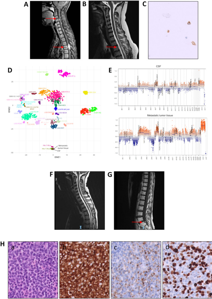
Pineoblastoma is a rare and aggressive malignancy that often affects pediatric populations. Accurate diagnosis is challenging due to histological overlap with other central nervous system tumors and limited molecular data. DNA methylation profiling and analysis of circulating tumor DNA (derived from both cell dissemination as well as cell-free- cfDNA) in cerebrospinal fluid (CSF) are emerging tools for precise tumor classification, in the field of pediatric central nervous system tumors. Here, we report a challenging case of a 17-year-old refugee girl with a previous diagnosis of a primitive neuroectodermal tumor. Formalin-fixed, paraffin-embedded tissue was not available for histopathological re-evaluation. However, the methylation profiling of low amount of CSF-derived DNA classified the tumor as "pineoblastoma, subtype miRNA processing altered 1, subclass A," enabling patient management. The diagnosis was later confirmed through tissue-based DNA methylation analysis of a secondary lesion, demonstrating that the epigenetic signature faithfully reflected tumor features. This case report highlights the potential of CSF-based DNA methylation profiling as a minimally invasive yet accurate diagnostic tool for pediatric CNS tumors. The concordance between CSF and tissue profiling supports the integration of liquid biopsy into diagnostic workflows, allowing for earlier diagnosis and personalized treatment strategies. However, more studies are needed to demonstrate the reliability of our approach in other CNS malignancies.
Keywords: Cerebrospinal fluid; DNA methylation; Diagnosis; Liquid biopsy; Pineoblastoma.
© 2025. The Author(s).
Conflict of interest statement
Declarations. Ethics approval and consent to participate: Appropriate intuitional approval and written consent was obtained from the patient’s family for this study. Consent for publication: Not applicable. Competing interests: The authors declare no competing interests.
Figures
References
-
- Cavalheiro S, Valsechi LC, Dastoli PA, Nicácio JM, Cappellano AM, Saba da Silva N et al (2023) Outcomes and surgical approaches for pineal region tumors in children: 30 years’ experience. J Neurosurg Pediatr 32:184–193 - PubMed
-
- Smith AB, Rushing EJ, Smirniotopoulos JG (2010) From the archives of the AFIP: lesions of the pineal region: radiologic-pathologic correlation. Radiographics 30:2001–2020 - PubMed
Publication types
MeSH terms
Grants and funding
LinkOut - more resources
Full Text Sources
Medical
