. 2025 Mar 8;23(1):127.
doi: 10.1186/s12964-025-02120-3.

IL-1β stimulates ADAMTS9 expression and contributes to preterm prelabor rupture of membranes

Affiliations

IL-1β stimulates ADAMTS9 expression and contributes to preterm prelabor rupture of membranes

Jiasong Cao et al. Cell Commun Signal. .

Abstract

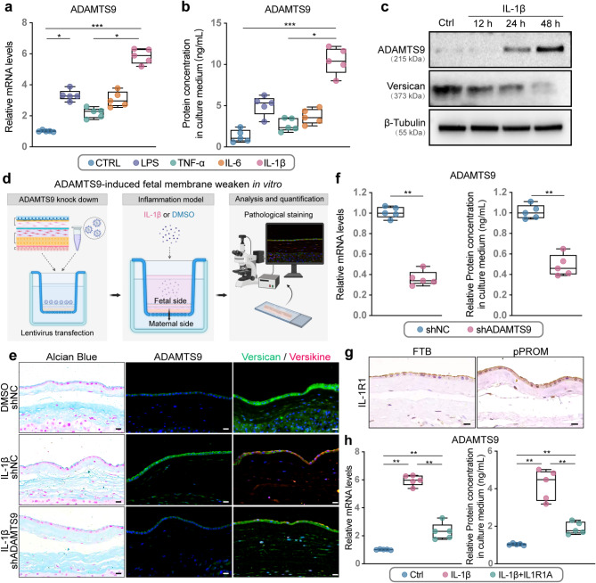
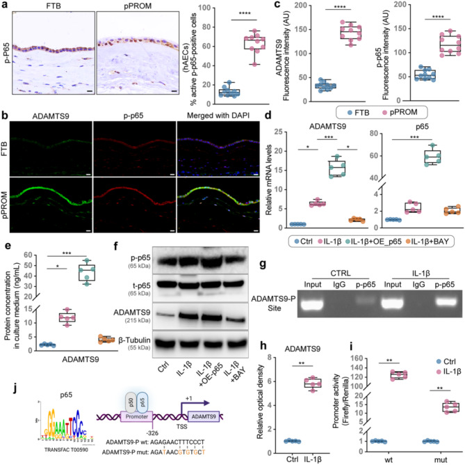
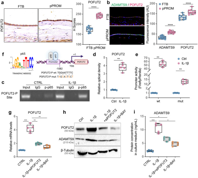
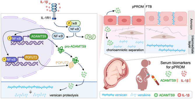
Background: Preterm prelabor rupture of membranes (pPROM) is a leading cause of neonatal morbidity and mortality. While intra-amniotic infection is a well-established driver of pPROM, the role of sterile intra-amniotic inflammation remains unclear. Recent evidence suggests that interleukin-1 beta (IL-1β) promotes extracellular matrix (ECM) remodeling via downstream effectors, a disintegrin-like and metalloproteinase domain with thrombospondin type 1 motif 9 (ADAMTS9), while protein O-fucosyltransferase 2 (POFUT2) facilitates its O-fucosylation and secretion, amplifying ECM degradation. This study investigates how IL-1β-triggered nuclear factor kappa-B (NF-κB) activation promotes ADAMTS9 and POFUT2 expression, ultimately driving fetal membrane ECM remodeling and weakening in pPROM without signs of intra-amniotic infection.

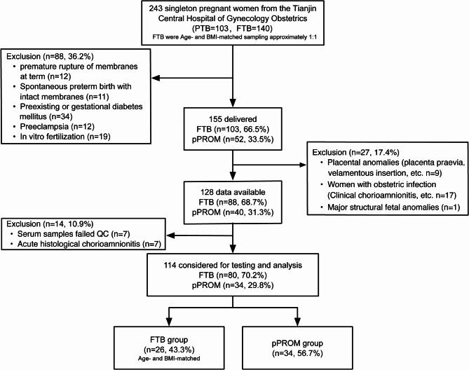
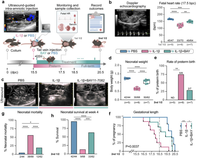
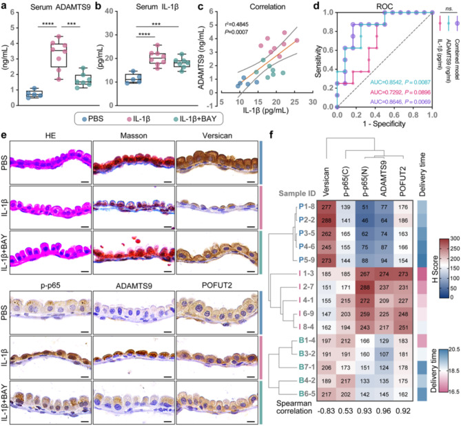
Methods: A nested case-control study included maternal serum and fetal membrane samples from 60 pregnant women (34 pPROM, 26 full-term births [FTB]). ELISA measured serum levels of IL-1β and ADAMTS9, and their correlations were analyzed. Mechanistic studies utilized primary human amniotic epithelial cells (hAECs) and fetal membrane-decidua explants with IL-1β treatment. The role of NF-κB was explored using chromatin immunoprecipitation (ChIP) and luciferase assays to assess NF-κB binding to the promoters of ADAMTS9 and POFUT2. A murine model of sterile intra-amniotic inflammation under ultrasound-guided IL-1β injection was used to validate in vitro findings and assess pregnancy outcomes.

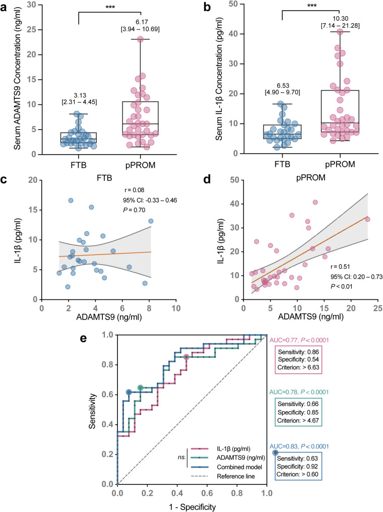
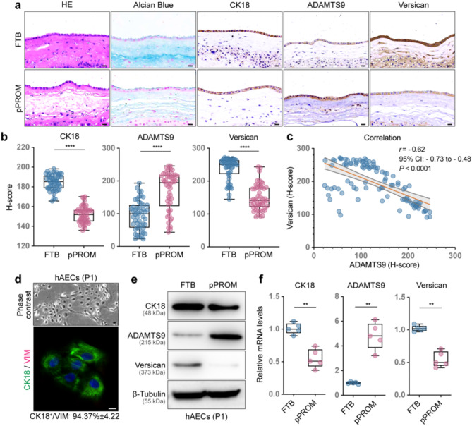
Results: Serum IL-1β and ADAMTS9 levels at 16 weeks of gestation were significantly higher in pPROM cases compared to FTB controls (P < 0.001). A combined model of these biomarkers demonstrated high predictive accuracy for pPROM (AUC = 0.83). Mechanistically, IL-1β activated NF-κB, leading to its binding to the promoters of ADAMTS9 and POFUT2. NF-κB activation promoted ADAMTS9 expression, while POFUT2 enhanced its secretion. Together, these processes drove versican degradation and ECM weakening. Intra-amniotic administration of IL-1β in mice induced fetal membrane weakening, preterm birth, and adverse neonatal outcomes, which were mitigated by the NF-κB inhibitor BAY 11-7082 treatment.

Conclusion: Maternal serum ADAMTS9 levels at mid-gestation are promising non-invasive biomarkers for pPROM risk stratification. Mechanistically, IL-1β-induced NF-κB activation promotes ADAMTS9 expression and POFUT2-dependent secretion, contributing to fetal membrane weakening. These findings provide new insights into the role and potential therapeutic target for sterile intra-amniotic inflammation in pPROM.

Keywords: ADAMTS9; Interleukin-1 beta; NF-kappa B; Preterm prelabor rupture of fetal membranes; Protein O-fucosyltransferase 2.


Conflict of interest statement

Declarations. Ethical approval: Upon the declaration of Helsinki, the clinical sample was approved by the Obstetrics and Gynecology Ethics Committee of Tianjin Central Hospital (Approval No. 2021KY113). The confidentiality of the patients was strictly protected, and no personal data were required for this study. All participants provided written informed consent. All animal procedures, including euthanasia, were carried out following ARRIVE guidelines and approved by the Institutional Review Board of Nankai University. Consent for publication: All authors have agreed to publish this manuscript. Competing interests: The authors declare no competing interests.

Figures

Fig. 1
Fig. 1
The patient flow diagram of the study population
Fig. 2
Fig. 2
The predictive efficacy of individual and composite biomarkers in predicting preterm prelabor rupture of membranes (pPROM). (a, b) Differences in prenatal serum IL-1β and ADAMTS9 concentrations between full-term birth (FTB; n = 26) and pPROM (n = 34) were determined using the Mann‒Whitney U test. (c, d) The correlation between serum IL-1β and ADAMTS9 in women with FTB and pPROM was evaluated using Spearman’s correlation coefficient (r). The orange line indicates the slope of the linear regression. The shadowed area indicates a 95% confidence interval, and the gray dashed line indicates the reference line. (e) Comparison of ROC curves of single and combined markers for predicting pPROM using the Delong method (n = 60). In the box-and-whisker plots, the midlines represent the medians, the boxes indicate the interquartile ranges, and the whiskers represent the minimum and maximum values. ***P < 0.001, ns. P > 0.05
Fig. 3
Fig. 3
The association between highly expressed ADAMTS9-induced versican reduction and fetal membrane weakening in pPROM. (a) Representative images of HE staining, Alcian staining, and immunohistochemical (IHC) staining of CK18, ADAMTS9, and versican in FTB and pPROM fetal membranes. Scale bars: 20 μm. (b) CK18, ADAMTS9, and versican expression were quantified using the H-score method in five random fields from FTB (n = 50) and pPROM (n = 50) fetal membranes (Mann‒Whitney U test). (c) The correlation between amniotic ADAMTS9 and versican expression in FTB and pPROM was assessed using Spearman’s correlation coefficient (r). The orange line indicates the slope of the linear regression. The shadowed area indicates a 95% confidence interval, and the gray dashed line indicates the reference line. (d) Brightfield image (top) and CK18/VIM expression (bottom) in primary hAECs. Scale bar: 10 μm. (e, f) CK18, ADAMTS9, and versican protein (e) and mRNA (f) expression in primary hAECs from FTB and pPROM (Mann‒Whitney U test). In the box-and-whisker plots, the midlines indicate the medians, the boxes indicate the interquartile ranges, and the whiskers indicate the minimum and maximum values. **P < 0.01, ****P < 0.0001
Fig. 4
Fig. 4
IL-1β signaling promotes chorioamniotic membrane separation in fetal membrane-decidual explants by upregulating hAEC-derived ADAMTS9 expression. (a. b) ADAMTS9 mRNA (left) and secreted protein (right) expression in hAECs after the indicated treatment. Kruskal‒Wallis and Dunn’s post hoc tests were used for comparisons between groups. (c) IL-1β-induced cytosolic ADAMTS9 and membrane versican protein expression in hAECs. (d) A schematic illustration of the IL-1β-induced fetal membrane weakening explant model. (e) Representative images of Alcian staining and immunofluorescence (IF) staining of ADAMTS9 and versican/versiline in explants after treatment with IL-1β alone or combined with shADAMTS9. (f) Left: ADAMTS9 mRNA expression in hAECs isolated from the explant model (Mann‒Whitney U test); Right: ADAMTS9 concentration in culture medium obtained from the explant model was tested by ELISA (Mann‒Whitney U test). (g) Immunostaining of IL-1R1 in hAECs from the fetal membrane of FTB and pPROM. (h) ADAMTS9 mRNA expression in hAECs (left) and ADAMTS9 concentration in culture medium (right) after treatment with IL-1β alone or in combination with IL-1R1A (Kruskal‒Wallis test with Dunn’s post hoc multiple comparisons). Scale bar: 20 μm. In the box-and-whisker plots, the midlines indicate the medians, the boxes indicate the interquartile ranges, and the whiskers indicate the minimum and maximum values. *P < 0.05, **P < 0.01, ***P < 0.001, ****P < 0.0001
Fig. 5
Fig. 5
hAECs-derived ADAMTS9 expression is driven by NF-κB-mediated promoter activation. (a) IHC analysis of p-p65 expression in hAECs from FTB (n = 10) and pPROM (n = 10) (Mann‒Whitney U test). (b, c) Representative IF staining and semiquantitative analysis for ADAMTS9 (green) and p-p65 (red) in hAECs from FTB and pPROM (Mann‒Whitney U test). (d) ADAMTS9 and p65 mRNA expression in hAECs. (e) Quantification of ADAMTS9 protein secreted into the cultured medium after the indicated treatments using ELISA (Kruskal‒Wallis test with Dunn’s post hoc multiple comparisons). (f) Protein expression of p-p65, total-p65 (t-p65), and ADAMTS9 in hAECs after the indicated treatments. (g, h) Chromatin immunoprecipitation (ChIP) in combination with qPCR was used to detect p-p65 directly binding within the ADAMTS9 promoter region upon IL-1β treatment (Mann‒Whitney U test). (i) Dual luciferase reporter assay was conducted to measure the luciferase reporter activity of Luc-ADAMTS9-wild-type/mutant in hAECs treated with PBS or IL-1β (Mann‒Whitney U test). (j) A schematic diagram of the binding site between p-p65 and the ADAMTS9 promoter. Scale bar: 20 μm. In the box-and-whisker plots, the midlines indicate the medians, the boxes indicate the interquartile ranges, and the whiskers indicate the minimum and maximum values. *P < 0.05, **P < 0.01, ***P < 0.001, ****P < 0.0001
Fig. 6
Fig. 6
ADAMTS9 secretion is enhanced by NF-κB-mediated POFUT2 promoter activation and transcriptional regulation. (a) IHC staining and quantification of POFUT2 in hAECs of the fetal membrane (Mann‒Whitney U test). (b) IF staining of POFUT2 (red) and ADAMTS9 (green) in hAECs from the fetal membrane, with quantification based on fluorescence intensity (Mann‒Whitney U test). (c, d) ChIP, in combination with qPCR, was used to detect p-p65 directly binding within the POFUT2 promoter region upon IL-1β treatment (Mann‒Whitney U test). (e) A dual luciferase reporter assay was conducted to measure the luciferase reporter activity of Luc-POFUT2-wild-type/mutant in hAECs treated with PBS or IL-1β (Mann‒Whitney U test). (f) A schematic diagram of the binding site between p-p65 and the POFUT2 promoter. (g) POFUT2 mRNA expression in hAECs after the indicated treatments (Kruskal‒Wallis test with Dunn’s post hoc multiple comparisons). (h) Protein expression of POFUT2 and ADAMTS9 in hAECs after the indicated treatments. (i) Quantification of ADAMTS9 protein secreted into the cultured medium after the indicated treatments using ELISA (Kruskal‒Wallis test with Dunn’s post hoc multiple comparisons). Scale bar: 20 μm. In the box-and-whisker plots, the midlines indicate the medians, the boxes indicate the interquartile ranges, and the whiskers indicate the minimum and maximum values. *P < 0.05, **P < 0.01, ***P < 0.001, ****P < 0.0001
Fig. 7
Fig. 7
Targeting the NF-κB/ADAMTS9 signaling axis ameliorates sterile intra-amniotic inflammation-induced preterm labor and adverse outcomes. (a) Schematic illustration of the experimental strategy. (b) Representative Doppler echocardiography of the fetal heart (left) and fetal heart rate (right) at 17.5 days post coitum (dpc; Kruskal‒Wallis test with Dunn’s post hoc multiple comparisons). (c) Representative ultrasound images of the fetal mouse at 17.5 dpc. (d) Neonatal body weight (Fisher’s exact test). (e) Preterm birth rate (Kruskal‒Wallis test with Dunn’s post hoc multiple comparisons). (f) Gestational lengths were analyzed using the log-rank (Mantel-Cox) test. (g, h) Neonatal mortality and 4-week neonatal survival rates (Fisher’s exact test). In the box-and-whisker plots, the midlines indicate the medians, the boxes indicate the interquartile ranges, and the whiskers indicate the minimum and maximum values. *P < 0.05, **P < 0.01, ***P < 0.001, ****P < 0.0001
Fig. 8
Fig. 8
The prediction performance of individual and composite biomarkers in a murine PTB model. (a, b) Concentrations of ADAMTS9 and IL-1β in serum (Kruskal‒Wallis test with Dunn’s post hoc multiple comparisons). (c) Pearson correlation analysis for serum ADAMTS9 and IL-1β concentrations. (d) ROC analysis of ADAMTS9 and IL-1β alone or in combination to predict PTB in pregnant mice, with a pairwise comparison of AUROC, used the Delong method. (e) Representative images of HE, Masson, and IHC staining of versican, p-p65, ADAMTS9, and POFUT2 in mouse amniotic membrane tissues. Scale bar: 20 μm. (f) A heatmap depicting the H-score for versican, p-p65, ADAMTS9, and POFUT2 in mouse amniotic membrane tissues. In the box-and-whisker plots, the midlines indicate the medians, the boxes indicate the interquartile ranges, and the whiskers indicate the minimum and maximum values. *P < 0.05, **P < 0.01, ***P < 0.001, ****P < 0.0001
Fig. 9
Fig. 9
A graphical summary of potential predictive markers for pPROM and how IL-1β drives the occurrence and progression of pPROM. ADAMTS9 and IL-1β, particularly ADAMTS9, have potential clinical value as early predictive markers of pPROM. hAECs-derived ADAMTS9 plays a role in the cleavage of versican, leading to the separation, weakening, and eventual rupture of fetal membranes in pPROM. Mechanistically, aseptic inflammation-induced IL-1β/NF-κB pathway activation significantly contributes to upregulating POFUT2/ADAMTS9 expression in hAECs. This figure was created with BioRender.com

Similar articles

References

    1. Green ES, Arck PC. Pathogenesis of preterm birth: bidirectional inflammation in mother and fetus. Semin Immunopathol. 2020;42(4):413–29. - PMC - PubMed
    1. Liu Y, Gao L. Preterm labor, a syndrome attributed to the combination of external and internal factors. Maternal-Fetal Med. 2022;4(1):61–71.
    1. Ballabh P, de Vries LS. White matter injury in infants with intraventricular haemorrhage: mechanisms and therapies. Nat Rev Neurol. 2021;17(4):199–214. - PMC - PubMed
    1. Suff N, Story L, Shennan A. The prediction of preterm delivery: what is new? Semin Fetal Neonatal Med. 2019;24(1):27–32. - PubMed
    1. van der Ham DP, Vijgen SM, Nijhuis JG, van Beek JJ, Opmeer BC, Mulder AL, et al. Induction of labor versus expectant management in women with preterm prelabor rupture of membranes between 34 and 37 weeks: a randomized controlled trial. PLoS Med. 2012;9(4):e1001208. - PMC - PubMed

LinkOut - more resources