

Biological and biophysical properties of the tumor-localizing component of hematoporphyrin derivative
- PMID: 4005843
- PMCID: PMC4563995
Biological and biophysical properties of the tumor-localizing component of hematoporphyrin derivative
Abstract
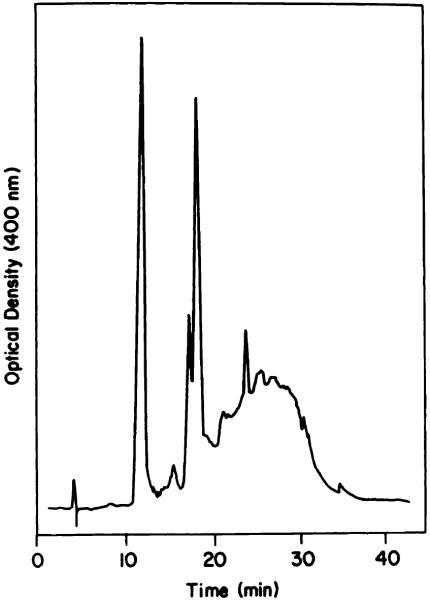
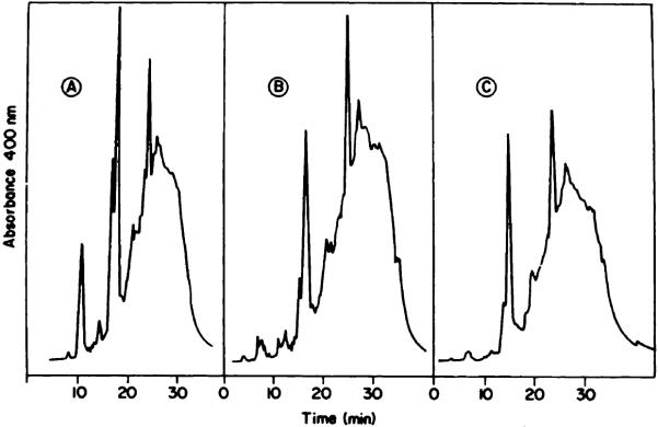
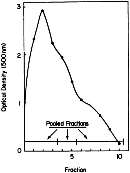
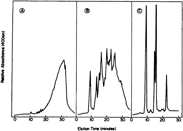
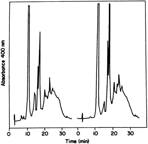
Reverse-phase chromatography, aqueous gel exclusion, and nonaqueous gel exclusion were assessed as procedures for preparative fractionation of the tumor-localizing product hematoporphyrin derivative. Porphyrin accumulation, fluorescence, and photodynamic cytotoxicity were monitored using the murine Sarcoma 180 tumor. Aqueous gel exclusion chromatography can provide a hematoporphyrin derivative fraction enriched in the tumor-localizing component. A further enrichment occurs when this procedure is carried out at 55 degrees C, but nonlocalizing porphyrins could not be eliminated. While providing a better separation, reverse-phase chromatography cannot provide a tumor-localizing fraction free from contaminating protoporphyrin. However, this and other contaminants can be eliminated from the tumor-localizing fraction via nonaqueous gel exclusion chromatography. This latter separation provides two tumor-localizing products: a fast-eluting fraction enriched in the major photosensitizing component(s); and a more complex slowly eluting fraction enriched in fluorescence localizers.
Figures

References
-
- Dougherty TJ, Boyle DG, Weishaupt KR, Henderson BA, Potter WR, Bellnier DA, Wityk KE. Photoradiation therapy—clinical and drug advances. In: Kessel D, Dougherty TJ, editors. Porphyrin Photosensitization. Plenum Publishing Corp; New York: 1983. pp. 3–13. - PubMed
-
- Kessel D. Hematoporphyrin and HPD: photophysics, photochemistry and Phototherapy. Photochem. Photobiol. 1984;39:851–859. - PubMed
-
- Spikes J. Photodynamic reactions in photomedicine. In: Regan JD, Parrish JA, editors. The Science of Photomedicine. Plenum Publishing Corp; New York: 1983. pp. 113–144.
-
- Gomer CJ, Dougherty TJ. Determination of [3H]- and [14C]hematoporphyrin derivative distribution in malignant and normal tissue. Cancer Res. 1979;39:146–151. - PubMed
Publication types
MeSH terms
Substances
Grants and funding
LinkOut - more resources
Full Text Sources
Other Literature Sources