. 2025 Mar 10;23(1):196.
doi: 10.1186/s12951-025-03243-y.

A bivalent spike-targeting nanobody with anti-sarbecovirus activity

Affiliations

A bivalent spike-targeting nanobody with anti-sarbecovirus activity

Iris C Swart et al. J Nanobiotechnology. .

Abstract

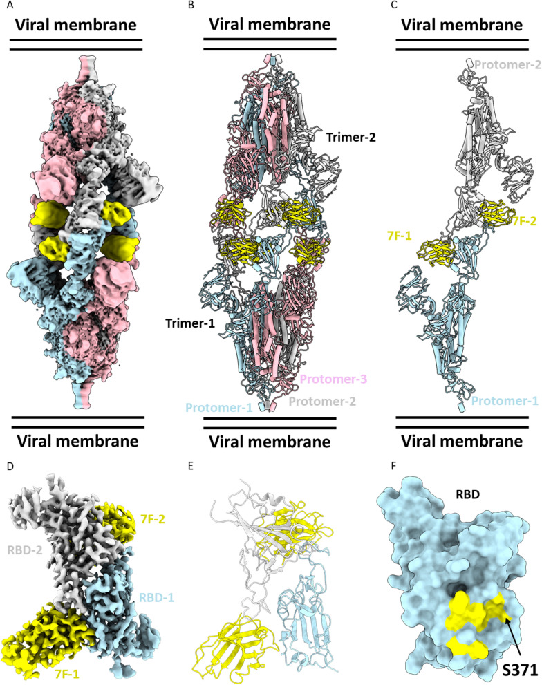
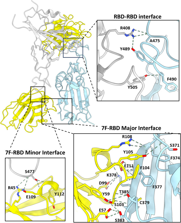
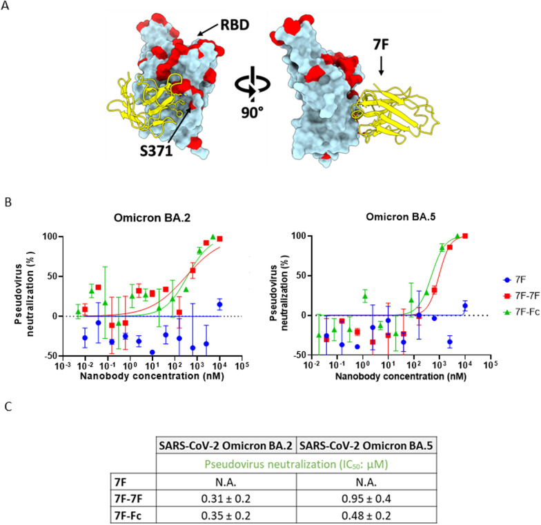
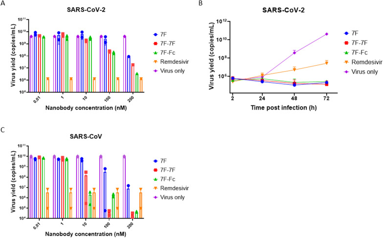
The continued emergence and zoonotic threat posed by coronaviruses highlight the urgent need for effective antiviral strategies with broad reactivity to counter new emerging strains. Nanobodies (or single-domain antibodies) are promising alternatives to traditional monoclonal antibodies, due to their small size, cost-effectiveness and ease of bioengineering. Here, we describe 7F, a llama-derived nanobody, targeting the spike receptor binding domain of sarbecoviruses and SARS-like coronaviruses. 7F demonstrates potent neutralization against SARS-CoV-2 and cross-neutralizing activity against SARS-CoV and SARS-like CoV WIV16 pseudoviruses. Structural analysis reveals 7F's ability to induce the formation of spike trimer dimers by engaging with two SARS-CoV-2 spike RBDs, targeting the highly conserved class IV region, though concentration dependent. Bivalent 7F constructs substantially enhance neutralization potency and breadth, up to more recent SARS-CoV-2 variants of concern. Furthermore, we demonstrate the therapeutic potential of bivalent 7F against SARS-CoV-2 in the fully differentiated 3D tissue cultures mirroring the epithelium of the human airway ex vivo. The broad sarbecovirus activity and distinctive structural features of bivalent 7F underscore its potential as promising antiviral against emerging and evolving sarbecoviruses.

Keywords: Cryo-EM; Nanobodies; Nanobody multimerization; Pandemic preparedness; SARS-CoV-2; Sarbecovirus; Spike trimer dimer formation.


Conflict of interest statement

Declarations. Ethics approval and consent to participate: Not applicable. Consent for publication: Not applicable. Competing interests: ID is an employee of Thermo Fisher Scientific. The remaining authors declare that they have no competing interests.

Figures

Fig. 1
Fig. 1
Camelid-derived nanobody 7F shows cross-reactivity activity against sarbecoviruses. A. ELISA-based reactivity of 7F to plate-immobilized spike ectodomain of SARS-CoV-2, SARS-CoV and RBD of SARS-like-CoV WIV16. Data points represent the mean ± SDM of n = 3 replicates from one representative of three independent experiments. B. Neutralization of SARS-CoV-2, SARS-CoV and SARS-like CoV WIV16 pseudovirus by serial diluted nanobody on VeroE6 cells. Data points represent the mean ± SDM of n = 3 replicates from one representative of two independent experiments. C. Schematic representation of the SARS-CoV-2 spike protein, with NTD and RBD labeled D. Binding of 7F to different spike domains analyzed by BLI. 7F was immobilized on NTA sensors, after which 7F was saturated in binding with either SARS-CoV-2 NTD or RBD. Data shown is based on a single experiment
Fig. 2
Fig. 2
Structural analysis of 7F in complex with the SARS-CoV-2 spike trimer. A. EM density map. The complex comprises six 7F molecules bound to two SARS-CoV-2 spike trimeric ectodomains. B. Atomic model of the six 7F molecules bound to two SARS-CoV-2 spike trimeric ectodomains. Spike protein protomers are in blue, grey, and pink, respectively. The 7F molecules are in yellow. C. Atomic model of a single protomer of each spike trimer, bound to two 7F molecules. Protomer-1 is blue, protomer-2 is grey and both 7F molecules are represented in yellow. D. Locally refined EM density map of the interaction site between two 7F molecules and two SARS-CoV-2 RBDs. E. Atomic model of the interaction site. RBD-1 is blue, RBD-2 is grey and 7F molecules are represented in yellow. F. Surface representation atomic model of SARS-CoV-2 RBD (blue) with the 7F epitope highlighted (yellow)
Fig. 3
Fig. 3
Functional and structural exploration of role of 7F in spike ACE2 interaction A. ELISA-based receptor binding inhibition assay. SARS-CoV-2 spike ectodomain preincubated with serially diluted 7F or control mAbs 87G7 (ACE2 binding competitor) and 47D11 (not competing with ACE2 binding), were added to a plate coated with soluble human ACE2. The spike-ACE2 interaction was quantified using HRP-conjugated antibody targeting the C-terminal Strep-tag fused to SARS-CoV-2 spike ectodomain. Data points represent the mean ± SDM of n = 3 replicates from one representative of three independent experiments. Concentration displayed in µg/mL corresponds to range of 0.001–1000 nM. B. Superimposition of human ACE2/SARS-CoV-2 complex (PDB: 6VW1) locally refined model of SARS-CoV-2 RBD-7F complex protein structure
Fig. 4
Fig. 4
Structural basis for sdAb-7F binding to the SARS-CoV-2 RBD. Ribbon representation of the dimeric RBD-7F complex, with inset panels showing detailed interactions for the major and minor interfaces
Fig. 5
Fig. 5
Enhanced neutralization potency of bivalent 7F constructs A. Mapping of sarbecovirus amino acid conservation onto the surface representation of SARS-CoV-2 RBD in complex with 7F. For comparison, residues 1 to 84 of the RBD-bound ACE2 (PDB ID: 6M0J) are shown as a silhouette. B. Design of different 7F constructs, from left to right: 7F monomer, genetically linked bivalent 7F-7F and 7F-Fc fusion. C. ELISA binding curves showing nanobody binding to immobilized SARS-CoV-2 spike ectodomain (left panel), SARS-CoV spike ectodomain (middle panel) and SARS-like CoV WIV16 RBD (right panel). Data points represent the mean ± SDM, for n = 3 replicates from one representative of three independent experiments. D. Nanobody mediated neutralization of luciferase encoding VSV particles pseudotyped with spike proteins of (left panel to right panel) SARS-CoV-2, SARS-CoV and SARS-like CoV WIV16 on VeroE6 cells. Data points represent the mean ± SDM, for n = 3 replicates from one representative of two independent experiments. E. Table showing the ELISA-based apparent binding affinities (KD) and pseudovirus neutralization based IC50 values of the monovalent and bivalent 7F constructs against SARS-CoV-2, SARS-CoV and SARS-like CoV WIV16 proteins or pseudoviruses, respectively. IC50 and KD values (± standard deviation) were calculated from the binding and neutralization curves displayed in B and C, respectively
Fig. 6
Fig. 6
Dimerization increases the neutralization breadth of 7F against SARS-CoV-2 variants-of-concern. A. Mapping of SARS-CoV-2 amino acid conservation onto the surface of SARS-CoV-2 RBD in complex with 7F. B. Nanobody mediated neutralization of luciferase encoding VSV particles pseudotyped with spike proteins of (left) SARS-CoV-2 Omicron BA.2 and (right) SARS-CoV-2 Omicron BA.5 on VeroE6 cells. Data points represent the mean ± SDM, for n = 3 replicates from one representative of two independent experiments. C. Summary of the IC50 values of the monovalent and bivalent 7F constructs against SARS-CoV-2 Omicron BA.2 and BA.5 pseudoviruses. IC50 values (± standard deviation) were calculated from the neutralization curves displayed in B
Fig. 7
Fig. 7
Monovalent and bivalent 7F constructs neutralize authentic SARS-CoV-2 in A549 cells and HAE cell cultures and authentic SARS-CoV in Vero cells. A. Neutralization of SARS-CoV-2 in A549ACE2+TMPRSS2+ cells. Virus was pre-incubated with serial diluted nanobody, or 10 µM remdesivir, for 30 min before infecting A549ACE2+TMPRSS2+ cells. Infection was quantified by measuring the virus yield (viral RNA copies/ml, as determined with RT-qPCR) in cell culture supernatants of SARS-CoV-2 infected cells. B. Neutralization of SARS-CoV-2 in HAE cell culture. HAE cultures were incubated with SARS-CoV-2 and 100 nM nanobodies or 10 µM remdesivir on the apical side for 2 h. Nanobody incubation was repeated every 24 h. Graph showing the quantification of viral replication in the cultures, evaluated by RT-qPCR. C. Neutralization of SARS-CoV in Vero cells. Virus was pre-incubated with serial diluted nanobody, or 10 µM remdesivir, for 30 min before infecting Vero cells. Infection was quantified by measuring the virus yield (viral RNA copies/ml, as determined with RT-qPCR) in cell culture supernatants of SARS-CoV infected cells

References

    1. Zhong NS, Zheng BJ, Li YM, Poon N, Xie ZH, Chan KH, et al. Epidemiology and cause of severe acute respiratory syndrome (SARS) in Guangdong People’s Republic of China in February, 2003. Lancet. 2003;362(9393):1353–8. - PMC - PubMed
    1. Zhou P, Yang XL, Wang XG, Hu B, Zhang L, Zhang W, et al. A pneumonia outbreak associated with a new coronavirus of probable bat origin. Nature. 2020;579(7798):270–3. - PMC - PubMed
    1. Lu R, Zhao X, Li J, Niu P, Yang B, Wu H, et al. Genomic characterisation and epidemiology of 2019 novel coronavirus: implications for virus origins and receptor binding. The Lancet. 2020;395(10224):565–74. - PMC - PubMed
    1. Ge XY, Li JL, Yang XL, Chmura AA, Zhu G, Epstein JH, et al. Isolation and characterization of a bat SARS-like coronavirus that uses the ACE2 receptor. Nature. 2013;503(7477):535–8. - PMC - PubMed
    1. Li W, Moore MJ, Vasilieva N, Sui J, Wong SK, Berne MA, et al. Angiotensin-converting enzyme 2 is a functional receptor for the SARS coronavirus. Nature. 2003;426(6965):450–4. - PMC - PubMed

MeSH terms

LinkOut - more resources