. 2025 Apr;14(11):e2404186.
doi: 10.1002/adhm.202404186. Epub 2025 Mar 10.

Supramolecular Additive Screening to Engineer Microfibrous Rafts for Expansion of Pluripotent Stem Cells in Dynamic Suspension

Affiliations

Supramolecular Additive Screening to Engineer Microfibrous Rafts for Expansion of Pluripotent Stem Cells in Dynamic Suspension

Johnick F van Sprang et al. Adv Healthc Mater. 2025 Apr.

Abstract

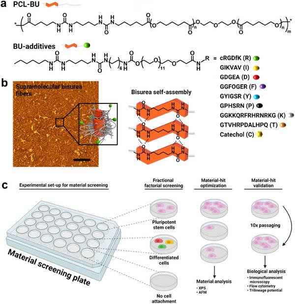
Human induced pluripotent stem cells (hiPSCs) hold the potential to generate any human tissue for transplantation in regenerative therapies. These complex cell therapies require billions of cells, which is challenging to acquire in planar adherent cultures. Transitioning hiPSCs to 3D suspension culture on microcarrier materials, often bead-shaped, improves the total surface area accessible to cells, thereby enabling culture scale-up. However, bead-shaped microcarriers do not have the optimal shape configuration, because it is the lowest surface-to-volume ratio of all geometrical shapes, and it also induces uncontrolled cell clumping. Application of synthetic, microfibrous rafts as a replacement for bead-shaped microcarriers potentially solves these issues. Here, microfibrous rafts are engineered by first screening a supramolecular biomaterial library composed of bisurea (BU)-peptide conjugate additives for its ability to induce hiPSC adhesion and maintenance of its pluripotent state, followed by electrospinning the screening-hit into raft-like structures. The resulting rafts contain cylinder-like microfibers, which have a higher surface-to-volume ratio compared to conventional bead-shaped microcarriers, and the flat configuration of the rafts prevents clumping.

Keywords: biomaterial; electrospinning; hiPSC; pluripotency; screening; supramolecular.


Conflict of interest statement

The authors declare no conflict of interest.

Figures

Figure 1
Figure 1
Schematic overview of the bisurea material library screening. a) Chemical structures of PCL‐BU and BU‐additives. The R‐group refers to the listed peptide sequences or catechol group, followed by a one‐letter code that is used as abbreviation. b) Atomic force phase micrograph of a PCL‐BU polymeric thin film depicting hard phase BU fiber‐like structures. To the right is a schematic representation of self‐assembly of BU moieties. Scale bar, 200 nm. c) A schematic overview of the bisurea materials screening and follow‐up experimental workflow.
Figure 2
Figure 2
Representative immunofluorescence microscopy images of hiPSCs on bisurea materials derived from the fractional factorial screening library after 3 days of culture. Black images are BU‐based materials on which no hiPSCs were found after 3 days of culture. Scale bars, 200 µm.
Figure 3
Figure 3
Fractional factorial screening output on hiPSC adhesion and pluripotency. a) The left graph shows the quantification of hiPSC count on bisurea materials after 3 days of culture. Data are represented as a mean ± s.e.m., N = 3. The right graph shows the effect size of single BU‐additives and two additive combinations. b) The left graph shows the quantification of hiPSC pluripotency on bisurea materials after 3 days of culture. Data are represented as a mean ± s.e.m., N = 3. The right graph shows the effect size of single BU‐additives and two additive combinations. c) Immunofluorescent microscopy images of hiPSCs on BU‐additive materials that had a significant effect size on pluripotency percentage. Scale bars, 200 µm. a,b) Statistical significance was attributed to values of p < 0.05 as determined by an effect normal plot. *p < 0.05, **p < 0.01, ***p < 0.001, ****p < 0.0001.
Figure 4
Figure 4
Screening‐hit optimization by variation of additive mol percentage. Top row shows immunofluorescent images of hiPSCs after a 3‐day culture period on bisurea materials with variations in additive concentration of the screening hit. Scale bars, 200 µm. Bottom row shows atomic force phase micrographs of PCL‐BU polymeric thin films with variations in additive concentration of the screening hits. Scale bars, 200 nm.
Figure 5
Figure 5
Material‐hit validation for hiPSC pluripotency after 35 days. a) A Schematic overview of the experimental workflow to assess long‐term pluripotency of hiPSCs on the bisurea material‐hit. b) Immunofluorescence microscopy images of hiPSCs after 4‐day culture period, staining transcription factors NANOG, OCT‐3/4, and SOX2. c) Immunofluorescence images of ectodermal cells after a 7‐day differentiation period, staining SOX1 and Otx2. d) Immunofluorescence images of mesodermal cells after a 5‐day differentiation period, staining HAND1 and Brachyury. e) Immunofluorescence images of endodermal cells after a 5‐day differentiation period, staining SOX17 and GATA4. b–e) Scale bars, 40 µm. f) Plot shows flow cytometry quantification of OCT‐3/4+ cells.
Figure 6
Figure 6
Bisurea‐based carrier platforms for expansion of hiPSCs. a) Top‐row depicts digital microscopy images of electrospun meshes after being cut into carrier platforms, middle row shows these carrier platforms after a 3‐day culture period. The bottom row shows SEM micrographs of the electrospun meshes. Scale bars, 10 µm. b–c) Immunofluorescence confocal microscopy images of hiPSCs after a 3‐day suspension culture period on carrier platform. Scale bars, 50 µm.

Similar articles

References

    1. Takahashi K., Yamanaka S., Cell 2006, 126, 663. - PubMed
    1. Okita K., Ichisaka T., Yamanaka S., Nature 2007, 448, 313. - PubMed
    1. Takahashi K., Tanabe K., Ohnuki M., Narita M., Ichisaka T., Tomoda K., Yamanaka S., Cell 2007, 131, 861. - PubMed
    1. Takebe T., Sekine K., Enomura M., Koike H., Kimura M., Ogaeri T., Zhang R. R., Ueno Y., Zheng Y. W., Koike N., Aoyama S., Adachi Y., Taniguchi H., Nature 2013, 499, 481. - PubMed
    1. Zeng Z., Huang B., Parvez R. K., Li Y., Chen J., Vonk A. C., Thornton M. E., Patel T., Rutledge E. A., Kim A. D., Yu J., Grubbs B. H., McMahon J. A., Pastor‐Soler N. M., Hallows K. R., McMahon A. P., Li Z., Nat. Commun. 2021, 12, 3641. - PMC - PubMed

LinkOut - more resources