. 2025 Mar 10;10(5):e181443.
doi: 10.1172/jci.insight.181443.

NAD+ prevents chronic kidney disease by activating renal tubular metabolism

Affiliations

NAD+ prevents chronic kidney disease by activating renal tubular metabolism

Bryce A Jones et al. JCI Insight. .

Abstract

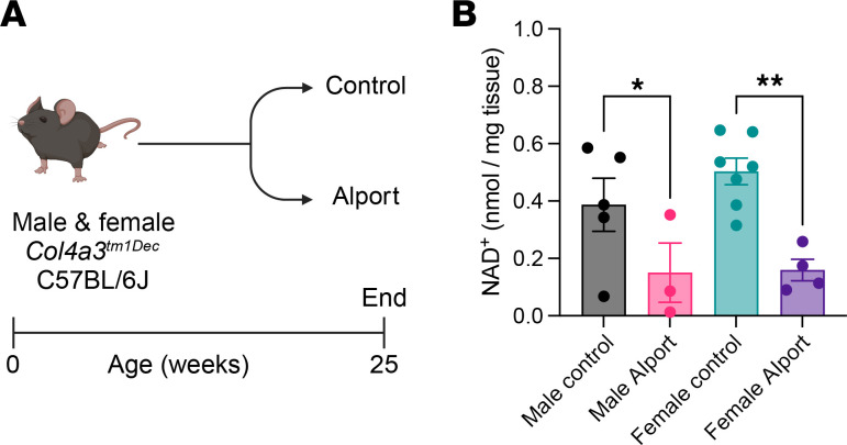
Chronic kidney disease (CKD) is associated with renal metabolic disturbances, including impaired fatty acid oxidation (FAO). Nicotinamide adenine dinucleotide (NAD+) is a small molecule that participates in hundreds of metabolism-related reactions. NAD+ levels are decreased in CKD, and NAD+ supplementation is protective. However, both the mechanism of how NAD+ supplementation protects from CKD, as well as the cell types involved, are poorly understood. Using a mouse model of Alport syndrome, we show that nicotinamide riboside (NR), an NAD+ precursor, stimulated renal PPARα signaling and restored FAO in the proximal tubules, thereby protecting from CKD in both sexes. Bulk RNA-sequencing showed that renal metabolic pathways were impaired in Alport mice and activated by NR in both sexes. These transcriptional changes were confirmed by orthogonal imaging techniques and biochemical assays. Single-nuclei RNA sequencing and spatial transcriptomics, both the first of their kind to our knowledge from Alport mice, showed that NAD+ supplementation restored FAO in proximal tubule cells. Finally, we also report, for the first time to our knowledge, sex differences at the transcriptional level in this Alport model. In summary, the data herein identify a nephroprotective mechanism of NAD+ supplementation in CKD, and they demonstrate that this benefit localizes to the proximal tubule cells.

Keywords: Chronic kidney disease; Molecular biology; Nephrology; Pharmacology.


Conflict of interest statement

Conflict of interest: BAJ discloses stock ownership in Allazo Health Corp. (New York, New York, USA).

Figures

Figure 1
Figure 1. Kidney NAD+ is reduced in Alport mice.
(A) Experimental design: Control and Alport mice of both sexes were sacrificed at 25 weeks of age. (B) Alport mice had lower levels of kidney NAD+ than control mice. Significance was determined by 1-way ANOVA with the Holm-Šídák correction for multiple comparisons. *P < 0.05, **P < 0.01. Data are expressed as the means ± SEM, and each datum represents 1 mouse.
Figure 2
Figure 2. NAD+ supplementation protects the kidney in Alport mice.
(A) Experimental design: Control and Alport mice of both sexes were treated with or without NR between 10 weeks and 25 weeks of age. (B) NR treatment reduced 24-hour urinary albumin excretion in Alport mice of both sexes. (C) Representative images of PSR-stained kidneys acquired with polarized light. Yellow-green-orange birefringence is highly specific for fibrosis. (D) Quantification of PSR-stained kidneys shows that NR treatment reduced renal fibrosis in both sexes. Significance was determined by 1-way ANOVA with the Holm-Šídák correction for multiple comparisons. Data are expressed as the means ± SEM, and each datum represents 1 mouse. *P < 0.05, **P < 0.01, ****P < 0.0001. NAD+, nicotinamide adenine dinucleotide; NR, nicotinamide riboside; PSR, Picrosirius red; Veh, vehicle.
Figure 3
Figure 3. NAD+ supplementation prevents both glomerular and tubular injury in Alport mice.
(A) Representative images of immunohistochemistry for p57kip2, followed by periodic acid–Schiff poststaining without hematoxylin counterstaining. Podocyte nuclei are stained brown. (BD) Quantification of p57kip2 immunostaining with PodoCount, a validated algorithm to analyze p57kip2-stained whole-slide images. Podocyte volumetric density (B) was reduced in Alport mice and restored by NR treatment in both sexes. Alport mice had podocyte nuclear hypertrophy (C) and glomerular hypertrophy (D) that was reduced by NR treatment in both sexes. (E and F) Immunoblots for KIM-1, a tubular injury marker, in male (E) and female (F) kidney homogenate. Ponceau S, a nonspecific protein stain, was used as a loading control. (G and H) Quantification of immunoblots (E and F) shows that KIM-1 is increased in Alport mice and reduced by NR treatment in male (G) and female (H) mice. Scale bars represent 100 μm. Significance was determined by 1-way ANOVA with the Holm-Šídák correction for multiple comparisons. Data are expressed as the means ± SEM. Each datum represents 1 glomerulus (BD) or 1 mouse (G and H). *P < 0.05, **P < 0.01, ***P < 0.001, ****P < 0.0001. KIM-1, kidney injury molecule-1; NAD+, nicotinamide adenine dinucleotide; No., number; NR, nicotinamide riboside; Podo. Vol. Den., podocyte volumetric density; PSR, Picrosirius red; Veh, vehicle.
Figure 4
Figure 4. NAD+ supplementation activates renal metabolism in male Alport mice.
Bulk kidney cortex RNA-Seq data from control and Alport mice, treated with or without NR, were analyzed (N = 4 mice per group). (A and B) GO biological processes that are both (A) reduced in vehicle-treated male Alport mice (vs. vehicle-treated male control mice) and (B) increased in NR-treated male Alport mice (vs. vehicle-treated male Alport mice) are shown. (C and D) KEGG pathways that are both (C) reduced in vehicle-treated male Alport mice (vs. vehicle-treated male control mice) and (D) increased in NR-treated male Alport mice (vs. vehicle-treated male Alport mice) are shown. (E and F) Transcription factor analyses suggest that the RXR/PPARα gene regulatory network is (E) inhibited in vehicle-treated male Alport mice (vs. vehicle-treated male control mice) and (F) activated in NR-treated male Alport mice (vs. vehicle-treated male Alport mice). Processes (A and B), pathways (C and D), and transcription factors (E and F) that are directly involved in fatty acid metabolism are highly enriched in all comparisons, emphasized by either dark blue (decreased) or dark red (increased). (G) Partial KEGG graph for the PPAR signaling pathway (KEGG Entry No. 03320). The left and right sides of each gene box represent data from vehicle-treated male Alport mice (vs. vehicle-treated male control mice) and NR-treated male Alport mice (vs. vehicle-treated male Alport mice), respectively. The subpathway that was the most restored by NR treatment was FAO. Alp, Alport; ChEA, ChIP Enrichment Analysis; Ctrl, control; FAO, fatty acid oxidation; GO, gene ontology; KEGG, Kyoto Encyclopedia of Genes and Genomes; M, male; NAD+, nicotinamide adenine dinucleotide; NR, nicotinamide riboside; RXR, retinoid X receptor; TF, transcription factor; Veh, vehicle.
Figure 5
Figure 5. NAD+ supplementation activates renal metabolism in control mice, not just in Alport mice.
Bulk kidney cortex RNA-Seq data from control and Alport mice, treated with or without NR, were analyzed (N = 4 mice per group). GO biological processes and KEGG pathways that were simultaneously upregulated in both NR-treated control mice (vs. vehicle-treated control mice) and NR-treated Alport mice (vs. vehicle-treated Alport mice) were identified. (A and B) GO biological processes that were increased in NR-treated control mice (vs. vehicle-treated control mice) in males (A) and females (B). (C and D) KEGG pathways that were increased in NR-treated control mice (vs. vehicle-treated control mice) in males (C) and females (D). These data (AD) are presented adjacent to the corresponding sex-matched comparisons from NR-treated Alport mice (vs. vehicle-treated Alport mice) in Supplemental Figures 15 and 16. (E and F) Transcription factor analyses suggest that the RXR/PPARα gene regulatory network is activated in NR-treated control mice (vs. vehicle-treated control mice) in both males (E) and females (F). Ctrl, control; F, female; GO, gene ontology; KEGG, Kyoto Encyclopedia of Genes and Genomes; M, male; NAD+, nicotinamide adenine dinucleotide; NR, nicotinamide riboside; RXR, retinoid X receptor; Veh, vehicle.
Figure 6
Figure 6. NAD+ supplementation activates renal fatty acid metabolism.
(A and B) NR treatment increased kidney PGC-1α in both control and Alport mice. (CF) Kidney CPT1α (C and E) and MCAD (D and F), key players in mitochondrial FAO, were reduced in Alport mice and restored by NR treatment. Ponceau S, a nonspecific protein stain, was used as a loading control. Significance was determined by 1-way ANOVA with the Holm-Šídák correction for multiple comparisons. Data are expressed as the means ± SEM. Each datum represents 1 mouse. *P < 0.05, **P < 0.01, ****P < 0.0001. CPT1α, carnitine palmitoyltransferase 1-α; FAO, fatty acid oxidation; MCAD, medium-chain acyl-coenzyme A dehydrogenase; NAD+, nicotinamide adenine dinucleotide; NR, nicotinamide riboside; PGC-1α, PPARγ coactivator 1-α; Veh, vehicle.
Figure 7
Figure 7. snRNA-Seq of control and Alport mice, with and without NAD+ supplementation.
(A) The snRNA-Seq reduction by UMAP of 49,488 nuclei in 30 clusters. (B) Proportion of nuclei per cluster by condition. Myofibroblasts and immune cells were upregulated in Alport mice. The effect was mitigated by NR treatment. (C) Differentially expressed genes in proximal tubule cells (PT_S1/S2) of NR-treated Alport mice (vs. vehicle-treated Alport mice). Mice treated with NR had reduced expression of immune signaling transcripts and increased expression of metabolism-related genes (e.g., Acot1, Ehhadh, and Insig1). (D) Enriched pathways between NR-treated Alport mice and vehicle-treated Alport mice in the proximal tubule cell. NR treatment significantly impacted translation (ribosome), metabolism, endocrine function, and PPAR signaling in the proximal tubule cell. Ctrl, control; NAD+, nicotinamide adenine dinucleotide; NR, nicotinamide riboside; UMAP, uniform manifold approximation and projection; Veh, vehicle. aPT, adaptive proximal tubules; B, B cell; CIC, cortical intercalated cells; CNT, connecting tubule; DCT, distal convoluted tubule; DendriC, dendritic cell; EC_1, endothelial cells 1; EC_2, endothelial cells 2; EC_Cycl, endothelial cells cycling; iDN, injury distal nephron; IMCL, intramedullary collecting cells; iPT, injury proximal tubules; MIC, medullary intercalated cells; MP_LP_1, macrophage or lymphocyte cells 1; MP_LP_2, macrophage or lymphocyte cells 2; NK_T, natural killer or T cell; PapEpi, papillary epithelial cells; PC, principal cell; POD, podocyte; PT_S1, proximal tubules segment 1; PT_S2, proximal tubules segment 2 cells; PT_S3, proximal tubules segment 3 cells 1; rDCT, regenerative distal convoluted tubule; sTAL, stressed thick ascending limb; STR_1, stromal cell 1; STR_2, stromal cell 2; STR_3, stromal cell 3; STR_4, stromal cell 4; TAL, thick ascending limb.
Figure 8
Figure 8. Spatial localization of Cpt1a and Acadm.
(A) Expression of mitochondrial transcription factor A (Tfam), carnitine palmitoyltransferase 1-α (Cpt1a), and medium-chain acyl-coenzyme A dehydrogenase (MCAD; Acadm) in cell types and conditions by snRNA-Seq. (B) Visium spatial transcriptomics was performed on vehicle-treated male control mice (left), vehicle-treated male Alport mice (center), and NR-treated male Alport mice (right). Cpt1a expression is depicted. (C) Cpt1a expression was restored with NR treatment. (D and E) Expression of Acadm was reduced in Alport mice and restored after NR treatment. (F) Differentially expressed genes within the cortical tubulointerstitium in NR-treated Alport mice (vs. vehicle-treated Alport mice). (G) Pathway enrichment in the cortical tubulointerstitium in NR-treated Alport mice (vs. vehicle-treated Alport mice). Avg., average; CI, confidence interval; Cortex, cortical tubulointerstitium; Ctrl, control; Glom, glomerulus; NAD+, nicotinamide adenine dinucleotide; NR, nicotinamide riboside; OR, odds ratio with 95% CI; Pct., percentage; Veh, vehicle. aPT, adaptive proximal tubules; B, B cell; CIC, cortical intercalated cells; CNT, connecting tubule; DCT, distal convoluted tubule; DendriC, dendritic cell; EC_1, endothelial cells 1; EC_2, endothelial cells 2; EC_Cycl, endothelial cells cycling; iDN, injury distal nephron; IMCL, intramedullary collecting cells; iPT, injury proximal tubules; MIC, medullary intercalated cells; MP_LP_1, macrophage or lymphocyte cells 1; MP_LP_2, macrophage or lymphocyte cells 2; NK_T, natural killer or T cell; PapEpi, papillary epithelial cells; PC, principal cell; POD, podocyte; PT_S1, proximal tubules segment 1; PT_S2, proximal tubules segment 2 cells; PT_S3, proximal tubules segment 3 cells 1; rDCT, regenerative distal convoluted tubule; sTAL, stressed thick ascending limb; STR_1, stromal cell 1; STR_2, stromal cell 2; STR_3, stromal cell 3; STR_4, stromal cell 4; TAL, thick ascending limb.

Update of

References

    1. Romagnani P, et al. Chronic kidney disease. Nat Rev Dis Primers. 2017;3:17088. doi: 10.1038/nrdp.2017.88. - DOI - PubMed
    1. Ralto KM, et al. NAD+ homeostasis in renal health and disease. Nat Rev Nephrol. 2020;16(2):99–111. doi: 10.1038/s41581-019-0216-6. - DOI - PMC - PubMed
    1. Poyan Mehr A, et al. De novo NAD+ biosynthetic impairment in acute kidney injury in humans. Nat Med. 2018;24(9):1351–1359. doi: 10.1038/s41591-018-0138-z. - DOI - PMC - PubMed
    1. Clark AJ, et al. The significance of NAD+ biosynthesis alterations in acute kidney injury. Semin Nephrol. 2022;42(3):151287. doi: 10.1016/j.semnephrol.2022.10.013. - DOI - PMC - PubMed
    1. Tran MT, et al. PGC1α drives NAD biosynthesis linking oxidative metabolism to renal protection. Nature. 2016;531(7595):528–532. doi: 10.1038/nature17184. - DOI - PMC - PubMed

MeSH terms