[Preprint]. 2025 May 23:2025.02.27.637541.
doi: 10.1101/2025.02.27.637541.

Pediatric cerebrospinal fluid immune profiling distinguishes pediatric-onset multiple sclerosis from other pediatric-onset acute neurological disorders

Affiliations

Pediatric cerebrospinal fluid immune profiling distinguishes pediatric-onset multiple sclerosis from other pediatric-onset acute neurological disorders

Diego A Espinoza et al. bioRxiv. .

Abstract

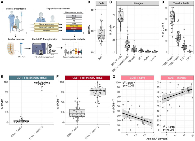
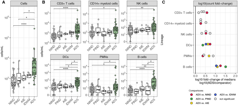
The cerebrospinal fluid (CSF) provides a unique glimpse into the central nervous system (CNS) compartment and offers insights into immune processes associated with both healthy immune surveillance as well as inflammatory disorders of the CNS. The latter include demyelinating disorders, such as multiple sclerosis (MS) and myelin oligodendrocyte glycoprotein antibody-associated disease (MOGAD), that warrant different therapeutic approaches yet are not always straightforward to distinguish on clinical and imaging grounds alone. Here, we establish a comprehensive phenotypic landscape of the pediatric CSF immune compartment across a range of non-inflammatory and inflammatory neurological disorders, with a focus on better elucidating CNS-associated immune mechanisms potentially involved in, and discriminating between, pediatric-onset MS (MS) and other pediatric-onset suspected neuroimmune disorders, including MOGAD. We find that CSF from pediatric patients with non-inflammatory neurological disorders is primarily composed of non-activated CD4+ T cells, with few if any B cells present. CSF from pediatric patients with acquired inflammatory demyelinating disorders is characterized by increased numbers of B cells compared to CSF of both patients with other inflammatory or non-inflammatory conditions. Certain features, including particular increased frequencies of antibody-secreting cells (ASCs) and decreased frequencies of CD14+ myeloid cells, distinguish MS from MOGAD and other acquired inflammatory demyelinating disorders.


Conflict of interest statement

COMPETING INTERESTS The authors declare no conflict of interest related to this study.

Figures

Figure 1:
Figure 1:. Pediatric NIND CSF is primarily composed of memory CD4+ and CD8+ T cells, with an age-associated increase in CD8+ memory T cell frequency
(A) Schematic of CSF sample procurement and flow cytometry data acquisition. (B) Cell count range (cells/mL) of NIND CSF (n=33). (C) Frequencies of CD3+ T cells, CD14+ myeloid cells, NK cells, DCs, PMNs, and B cells in NIND CSF. (D) Frequencies of T-cell subsets, as percent of CD3+ T cells, in NIND CSF. (E) Frequencies of CD4+ naive and memory T cells, as percent of CD4+ T cells, in NIND CSF. (F) Frequencies of CD8+ naive and memory T cells, as percent of CD8+ T cells, in NIND CSF and (G) across the NIND age span.
Figure 2:
Figure 2:. An increase in CSF B-cell counts best differentiates ADS from both other inflammatory and non-inflammatory neurological disease cohorts
(A) Cell counts (cells/mL) in NIND CSF (n=33), PIND CSF (n=7), AIE CSF (n=6), IDWM CSF (n=4), and ADS CSF (n=35). (B) Cell counts (cells/mL) for CD3+ T cells, CD14+ myeloid cells, NK cells, DCs, PMNs, and B cells across the same cohorts as A. (C) log10 of the fold change of median cell counts, comparing the median cell count in ADS to the median cell count in other cohorts for each lineage. For Fig. 2A–B, Wilcoxon-rank-sum test used to compare ADS to NIND, PIND, AIE, and IDWM independently (* = p < 0.05, ** = p < 0.01, *** = p < 0.001, **** = p < 0.0001). NIND = non-inflammatory neurological disease, PIND = peripheral inflammatory neurological disease, AIE = autoimmune encephalitidies, IDWM = inherited disorders of white matter, ADS = acquired demyelinating syndromes.
Figure 3:
Figure 3:. Pediatric MS CSF exhibits increased ASC frequencies and decreased CD14+ myeloid cell frequencies when compared to MOGAD and other ADS.
(A) B-cell, (B) ASC, and (C) CD14+ myeloid cell frequencies and counts (cells/mL) in NIND CSF (n=33), PIND CSF (n=7), AIE CSF (n=6), IDWM CSF (n=4), other ADS CSF (n=10), MOGAD CSF (n=10), and MS CSF (n=15). (D) log10 of the fold change of median frequencies, comparing the median frequency in MS to the median frequency in other ADS and MOGAD for each cell population (lineage or subset). For Fig. 3A–C, Wilcoxon-rank-sum test used to compare MS to MOGAD, MS to other ADS, and MOGAD to other ADS (* = p < 0.05, ** = p < 0.01, *** = p < 0.001, **** = p < 0.0001). ASC = antibody secreting cell, NIND = non-inflammatory neurological disease, PIND = peripheral inflammatory neurological disease, AIE = autoimmune encephalitidies, IDWM = inherited disorders of white matter, other ADS = non-MS/non-MOGAD acquired demyelinating syndromes, MOGAD = myelin oligodendrocyte glycoprotein antibody-associated disease, MS = multiple sclerosis.
Figure 4:
Figure 4:. Ratio of CSF ASC to CSF CD14+ myeloid cell frequencies differentiates MS from MOGAD and other ADS
(A) Ratio of ASC frequencies to CD14+ myeloid cell frequencies (AMR) and (B) CSF-only neuroinflammatory composite scores (coNCS) across NIND CSF (n=33), PIND CSF (n=7), AIE CSF (n=6), IDWM CSF (n=4), and other ADS CSF (n=10), MOGAD CSF (n=10), and MS CSF (n=15). (C) Receiver operating characteristic (ROC) curves built for AMR or coNCS classifiers for the MS-vs-MOGAD/other ADS comparison, MS-vs-MOGAD comparison, and MS-vs-other ADS comparison, along with each corresponding area under the curve (AUC). For Fig. 4A–B, Wilcoxon-rank-sum used to compare MS to MOGAD, MS to other ADS, and MOGAD to other ADS (* = p < 0.05, ** = p < 0.01, *** = p < 0.001, **** = p < 0.0001). AMR = ASC to CD14+ myeloid cell ratio, coNCS = CSF-only neuroinflammatory composite score, NIND = non-inflammatory neurological disease, PIND = peripheral inflammatory neurological disease, AIE = autoimmune encephalitidies, IDWM = inherited disorders of white matter, other ADS = non-MS/non-MOGAD acquired demyelinating syndromes, MOGAD = myelin oligodendrocyte glycoprotein antibody-associated disease, MS = multiple sclerosis.

References

    1. Ousman SS, Kubes P. Immune surveillance in the central nervous system. Nat Neurosci. 2012;15(8):1096–1101. doi: 10.1038/nn.3161 - DOI - PMC - PubMed
    1. Stangel M, Fredrikson S, Meinl E, Petzold A, Stüve O, Tumani H. The utility of cerebrospinal fluid analysis in patients with multiple sclerosis. Nat Rev Neurol. 2013;9(5):267–276. doi: 10.1038/nrneurol.2013.41 - DOI - PubMed
    1. Ostkamp P, Deffner M, Schulte-Mecklenbeck A, et al. A single-cell analysis framework allows for characterization of CSF leukocytes and their tissue of origin in multiple sclerosis. Science Translational Medicine. 2022;14(673):eadc9778. doi: 10.1126/scitranslmed.adc9778 - DOI - PubMed
    1. Schafflick D, Xu CA, Hartlehnert M, et al. Integrated single cell analysis of blood and cerebrospinal fluid leukocytes in multiple sclerosis. Nature Communications. 2020;11(1):247. doi: 10.1038/s41467-019-14118-w - DOI - PMC - PubMed
    1. Han S, Lin YC, Wu T, et al. Comprehensive Immunophenotyping of Cerebrospinal Fluid Cells in Patients with Neuroimmunological Diseases. The Journal of Immunology. 2014;192(6):2551–2563. doi: 10.4049/jimmunol.1302884 - DOI - PMC - PubMed

Publication types