[Preprint]. 2025 Feb 27:2025.02.21.639315.
doi: 10.1101/2025.02.21.639315.

Distal mutations enhance catalysis in designed enzymes by facilitating substrate binding and product release

Affiliations

Distal mutations enhance catalysis in designed enzymes by facilitating substrate binding and product release

Niayesh Zarifi et al. bioRxiv. .

Update in

Abstract

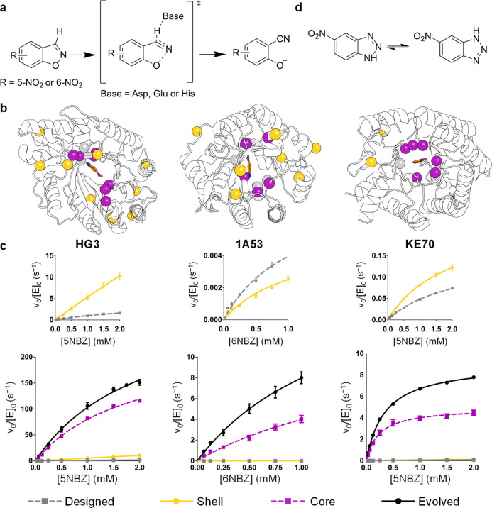
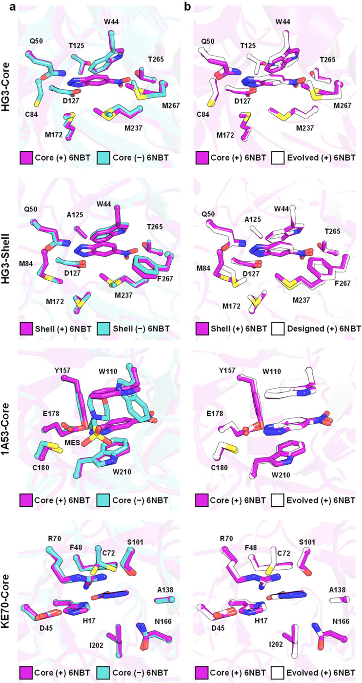
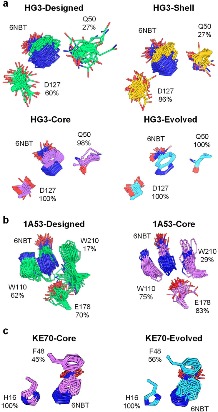
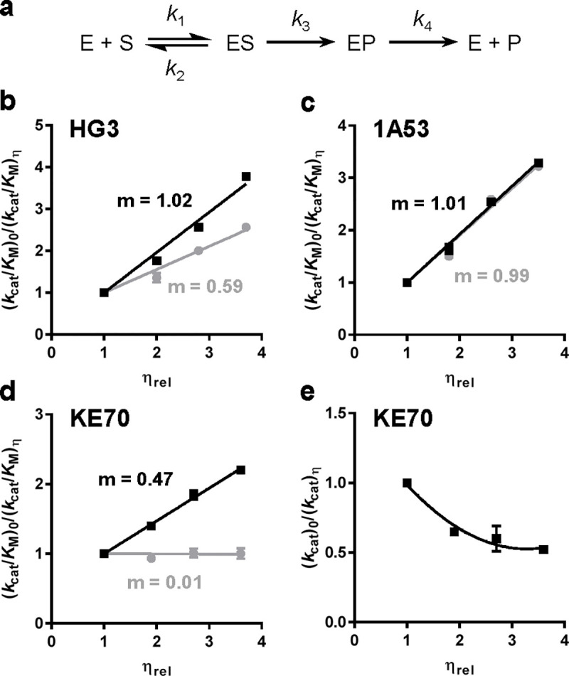
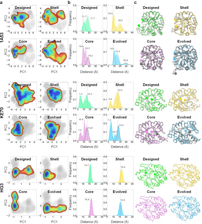
The role of amino-acid residues distant from an enzyme's active site in facilitating the complete catalytic cycle-including substrate binding, chemical transformation, and product release-remains poorly understood. Here, we investigate how distal mutations promote the catalytic cycle by engineering mutants of three de novo Kemp eliminases containing either active-site or distal mutations identified through directed evolution. Kinetic analyses, X-ray crystallography, and molecular dynamics simulations reveal that while active-site mutations create preorganized catalytic sites for efficient chemical transformation, distal mutations enhance catalysis by facilitating substrate binding and product release through tuning structural dynamics to widen the active-site entrance and reorganize surface loops. These distinct contributions work synergistically to improve overall activity, demonstrating that a well-organized active site, though necessary, is not sufficient for optimal catalysis. Our findings reveal critical roles that distal residues play in shaping the catalytic cycle to enhance efficiency, yielding valuable insights for enzyme design.

Keywords: Enzyme catalysis; X-ray crystallography; enzyme design; enzyme engineering; enzyme mechanisms; molecular dynamics.


Conflict of interest statement

Competing Interests The authors declare no competing interests.

Figures

Figure 1.
Figure 1.. Core and Shell variants of Kemp eliminases.
(a) Kemp eliminases catalyze the concerted deprotonation and ring opening of benzisoxazoles using an Asp, Glu or His catalytic base. (b) Core and Shell variants from the HG3, 1A53 and KE70 families contain either active-site (purple) or distal (yellow) mutations found by directed evolution, respectively. Mutations are listed on Supplementary Table 2. Catalytic residues are shown as grey spheres. (c) Michaelis–Menten plots of normalized initial rates as a function of substrate concentration are shown. Data represent the average of six or nine individual replicate measurements from two or three independent protein batches, with error bars indicating the SEM (mean ± SEM). (d) The 6-nitrobenzotriazole transition-state analogue tautomerizes in water, which allows it to bind to enzymes that react with either 5- or 6-nitrobenzisoxazole.
Figure 2.
Figure 2.. Crystal structures.
(a) The active sites of HG3-Core, HG3-Shell and KE70-Core do not change substantially upon binding of the 6-nitrobenzotriazole (6NBT) transition-state analogue. In 1A53-Core, a 2-(N-morpholino)ethanesulfonic acid (MES) molecule from the crystallization buffer occupies the 6NBT binding site, inducing a conformational change in W110. (b) Core variants display active-site configurations nearly identical to those of their Evolved counterparts, while HG3-Shell displays a configuration similar to HG3-Designed. The 1A53–2.5 crystal structure (PDB ID: 6NW4) was used for comparison with 1A53-Core, as 1A53-Evolved was unavailable. We re-refined the KE70-Evolved crystal structure with bound 6NBT (PDB ID: 3Q2D) to model missing residues (22–25 in chain A and 3–25 in chain B) into the available density and to flip 6NBT into a productive binding pose. In all cases, only the major conformer of each first-shell active-site residue is shown, with the 6NBT transition-state analogue at the center.
Figure 3.
Figure 3.. Ensemble refinement of Kemp eliminases.
Ensemble refinements of 6NBT-bound crystal structures for (a) HG3, (b) 1A53, and (c) KE70 variants reveal that distal and active-site mutations increase the proportion of productive substates in the conformational ensemble. Percentages next to each residue indicate the fraction of substates that form hydrogen bonds or π-stacking interactions with 6NBT.
Figure 4.
Figure 4.. Kinetic solvent viscosity effects.
(a) Kemp elimination reaction scheme. (b-e) Grey and black lines correspond to Core and Evolved variants, respectively. Kinetic solvent viscosity effects on kcat/KM for (a) HG3, (b) 1A53 and (c) KE70 variants show a linear dependence on relative viscosity (ηrel). The slope (m) corresponds to k3/k2+k3, where k2 is the rate constant of substrate dissociation and k3 is the rate constant for the chemical transformation. A slope of 1 indicates that k3 is much larger than k2m=k3/k3. A slope of 0 indicates that k3 is much smaller than k2m=k3/k2=0. (e) Kinetic solvent viscosity effects on kcat for KE70-Evolved show an inverse hyperbolic pattern, which is consistent with the presence of a solvent-sensitive internal isomerization of the enzyme-product complex. Data represent the average of 6 individual replicates from 2 independent protein batches, with error bars reporting the SEM.
Figure 5.
Figure 5.. Conformational landscapes of Kemp eliminases analyzed by molecular dynamics.
(a) Representation of the molecular dynamics trajectories projected into the two most important principal components (PC1, PC2) based on Cα contacts for each enzyme family. Color coding represents the relative population density of the states, with blue indicating the most populated wells and red representing the least populated regions. Details on conformational changes contributing to PC1 and PC2 are shown on Supplementary Figure 11. (b) Histograms of loop distances across the entire energy landscape for KE70 and 1A53 enzymes, or from the most populated well for HG3 enzymes. Loop distances were calculated between Cα carbons of residues 21 and 78 for KE70 variants, 58 and 188 for 1A53 variants, and 90 and 275 for HG3 variants (shown as spheres in panel c). The dashed line represents the average loop distance in crystal structures of each enzyme family. The multimodal distributions were deconvoluted using a Gaussian mixture model, with the median loop distances for each conformational state indicated in Å. (c) Representative snapshots for each conformational state sampled by the enzymes. These snapshots display loop distances equivalent to the median value for each population in the distribution. The Cα carbons of residues used to calculate loop distances are shown as spheres.

References

    1. Rauwerdink A. & Kazlauskas R.J. How the Same Core Catalytic Machinery Catalyzes 17 Different Reactions: the Serine-Histidine-Aspartate Catalytic Triad of α/β-Hydrolase Fold Enzymes. ACS Catalysis 5, 6153–6176 (2015). - PMC - PubMed
    1. Du S. et al. Conformational ensembles reveal the origins of serine protease catalysis. Science 387, eado5068 (2025). - PubMed
    1. Khersonsky O. et al. Optimization of the In-Silico-Designed Kemp Eliminase KE70 by Computational Design and Directed Evolution. Journal of Molecular Biology 407, 391–412 (2011). - PMC - PubMed
    1. Broom A. et al. Ensemble-based enzyme design can recapitulate the effects of laboratory directed evolution in silico. Nature Communications 11, 4808 (2020). - PMC - PubMed
    1. Otten R. et al. How directed evolution reshapes the energy landscape in an enzyme to boost catalysis. Science 370, 1442–1446 (2020). - PMC - PubMed

Publication types

LinkOut - more resources