[Preprint]. 2025 Feb 27:2025.02.26.640428.
doi: 10.1101/2025.02.26.640428.

Nanometer condensate organization in live cells derived from partitioning measurements

Affiliations

Nanometer condensate organization in live cells derived from partitioning measurements

Christina Dollinger et al. bioRxiv. .

Abstract

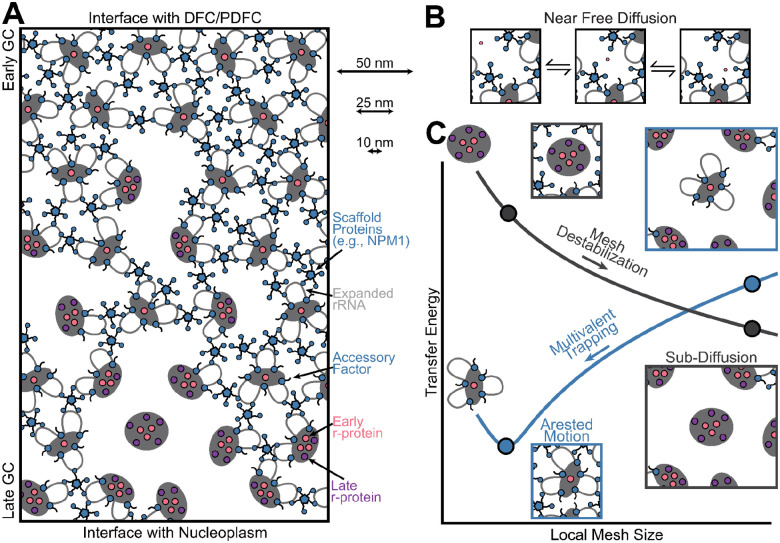
Biomolecules associate, forming condensates that house essential biochemical processes, including ribosome biogenesis. Unraveling how condensates shape macromolecular assembly and transport requires cellular measurements of nanoscale structure. Here, we determine the organization around and between specific proteins at nanometer resolution within condensates, deploying thermodynamic principles to interpret partitioning measurements of designed protein probes. When applied to the nucleolus as a proof of principle, the data reveals considerable inhomogeneity, deviating from that expected within a liquid-like phase. The inhomogeneity can be attributed to ribosome biogenesis, with the local meshwork weakening as biogenesis progresses, facilitating transport. Beyond introducing an innovative modality for biophysical interrogation, our results suggest condensates are far from uniform, simple liquids, a property we conjecture enables regulation and proofreading.


Conflict of interest statement

Competing interests: None

Figures

Figure 1:
Figure 1:. Local size exclusion measures the mesh size around a specific protein within a condensate.
(A) Standard state transfer free energy measurement requires the determination of the concentration, C, of a probe molecule between compartments. (B) Overexpression of probe, shown NPM1 C-terminal truncation with mGreenLantern (mGL), in U2OS with endogenous NPM1-mCherry. Np fixed sets the value of the nucleoplasm at the same LUTs dictated by a max partitioning of all images. (C) Quantification of the probe levels (bottom) and the endogenous NPM1 ΔGtr (top) between the GC and Np phases. Dashed lines show trends extrapolated to zero probe overexpression. Relative concentration units (RCUs) are the number of photons emitted at reference settings (methods). (D) Cells with low overexpression of indicated constructs. (E) Quantification of indicated EMG1 probes. (F) Transfer free energy measurement for a nucleolar protein without and with an N residue chain. (G) ΔGtr dependence on the number of chain residues added to the EMG1 probe. Calculation of the apparent mesh size shown (inset). Unless indicated, all images are Np fixed, and scale bars are 5 μm.
Figure 2:
Figure 2:. LSE reveals that the local mesh size increases during ribosome biogenesis.
(A) Schematic of ribosome biogenesis and the categories of proteins studied. (B) LSE for individual (top) and categorically grouped proteins (bottom, Fig S3). (C) Relationship between σ and ΔGint.tr. Fit assumes the ΔGint.tr is proportional to the mesh contact number density (σ3v, methods). (D) EM data showing the Np and GC separated by the dashed line (fire mode, top left). Image of segmented grains from EM image (top right). Graph of radial distribution function (RDF) from the center of the GC grains in EM images (bottom, Fig S5).
Figure 3:
Figure 3:. LDSC measures the distance between proteins.
(A) Linear breakdown of the ΔGtr for each NPM1 construct by its domains into energies as indicated. (B) Results of linear decomposition of the ΔGtr (Fig S6). (C) Schematic depicting the spatial coupling (Sp.Co.) as a function of chain length (top) and its calculation (bottom). (D) Length-dependent Sp.Co. (LDSC) between the N and C terminal domains of NPM1. Chain (red) of NPM1; not used in fit. (E) LDSC between EMG1 and NPM1. (F) LDSC between NPM1 and proteins grouped by role in ribosome biogenesis. For analytical form in D-F see methods.
Figure 4:
Figure 4:. LSE and LDSC provide a nanometer picture of molecular interactions and transport during ribosome biogenesis.
(A) Gradient of ribosome biogenesis and mesh sizes within the GC. Note that client molecules, including scaffold proteins that are in client-like states, are omitted to highlight mesh. (B) Depiction of free early r-protein transport within the GC meshwork. (C) Local mesh size dependence on transfer energy and transport for an early ribosomal intermediate (blue) and the completed ribosome (gray) due to local changes in the number of interactions (Fig S4, methods).

References

    1. Molliex A., Temirov J., Lee J., Coughlin M., Kanagaraj A. P., Kim H. J., Mittag T., Taylor J. P., Phase Separation by Low Complexity Domains Promotes Stress Granule Assembly and Drives Pathological Fibrillization. Cell 163, 123–133 (2015). - PMC - PubMed
    1. Feric M., Vaidya N., Harmon T. S., Mitrea D. M., Zhu L., Richardson T. M., Kriwacki R. W., Pappu R. V., Brangwynne C. P., Coexisting Liquid Phases Underlie Nucleolar Subcompartments. Cell 165, 1686–1697 (2016). - PMC - PubMed
    1. Brangwynne C. P., Eckmann C. R., Courson D. S., Rybarska A., Hoege C., Gharakhani J., Jülicher F., Hyman A. A., Germline P granules are liquid droplets that localize by controlled dissolution/condensation. Science 324, 1729–1732 (2009). - PubMed
    1. Mittag T., Pappu R. V., A conceptual framework for understanding phase separation and addressing open questions and challenges. Mol. Cell, doi: 10.1016/j.molcel.2022.05.018 (2022). - DOI - PMC - PubMed
    1. Musacchio A., On the role of phase separation in the biogenesis of membraneless compartments. EMBO J. 41, e109952 (2022). - PMC - PubMed

Publication types

LinkOut - more resources