[Preprint]. 2025 Feb 28:2025.02.28.640732.
doi: 10.1101/2025.02.28.640732.

The cerebrospinal fluid virome in people with HIV: links to neuroinflammation and cognition

Affiliations

The cerebrospinal fluid virome in people with HIV: links to neuroinflammation and cognition

Mattia Trunfio et al. bioRxiv. .

Abstract

Despite effective HIV suppression, neuroinflammation and neurocognitive issues are prevalent in people with HIV (PWH) yet poorly understood. HIV infection alters the human virome, and virome perturbations have been linked to neurocognitive issues in people without HIV. Once thought to be sterile, the cerebrospinal fluid (CSF) hosts a recently discovered virome, presenting an unexplored avenue for understanding brain and mental health in PWH. This cross-sectional study analyzed 85 CSF samples (74 from PWH on suppressive antiretroviral therapy, and 11 from controls without HIV, CWH) through shotgun metagenomics for DNA/RNA viruses. Taxonomic composition (reads and contigs), α and β diversity, and relative abundance (RA) of prokaryotic (PV), human eukaryotic (hEV), and non-human eukaryotic viruses (nhEV) were evaluated in relation to HIV infection, markers of neuroinflammation and neurodegeneration, cognitive functions, and depressive symptoms. Sensitivity analyses and post-hoc cluster analysis on the RA of viral groups and blood-brain barrier permeability were also performed. Of 46 read-positive CSF samples, 93.5% contained PV sequences, 47.8% hEV, and 45.6% nhEV. Alpha diversity was lower in PWH versus CWH, although p>0.05. At β diversity analysis, HIV status explained 3.3% of the variation in viral composition (p=0.016). Contigs retained 13 samples positive for 8 hEV, 2 nhEV, and 6 PV. Higher RA of PV was correlated with higher CSF S100β (p=0.002) and β-Amyloid 1-42 fragment (βA-42, p=0.026), while higher RA of nhEV with poorer cognitive performance (p=0.022). Conversely, higher RA of hEV correlated with better cognition (p=0.003) and lower βA-42 (p=0.012). Sensitivity analyses in virome-positive samples only confirmed these findings. Three CSF clusters were identified and showed differences in astrocytosis, βA-42, tau protein, and cognitive functions. Participants with hEV-enriched CSF showed better cognitive performance compared to those with virus-devoid and nhEV-enriched CSF (models'p<0.05). This study provides the first comprehensive description of the CSF virome in PWH, revealing associations with neuroinflammation and cognition. These findings highlight the potential involvement of the CSF virome in brain health and inform about its composition, origin, and potential clinical implications in people with and without HIV.

Keywords: Bacteriophages; Brain microbiome; Central nervous system; Cognitive functions; Depression; Eukaryotic viruses; HIV-associated neurocognitive disorders; Prokaryotic viruses.


Figures

Figure 1.
Figure 1.. Prevalence and relative abundance of CSF viral families by HIV status
Panels A and B show the prevalence of viral families in the CSF (proportion of samples positive for each viral taxon among all the CSF samples) of PWH and CWH, respectively. Three families were found in about half of CSF samples from both PWH and CWH: Siphoviridae and Myoviridae (double-stranded DNA phages belonging to the order of Caudovirales), and Genomoviridae (single-stranded DNA viruses). Panels C and D show the relative abundance (number of reads of the specific taxon over the total number of reads per sample) of the viral families in virome-positive samples from PWH and CWH, respectively. Myoviridae and Siphoviridae represented the 60.2% of the total viral genetic material detected in the CSF of PWH, followed by Herpesviridae (13.6%, human double-stranded DNA viruses), Podoviridae (10.4%, double-stranded DNA phages belonging to the order of Caudovirales), Virgaviridae (single-stranded RNA viruses of plants), and 18 other viral families at much lower proportions. Siphoviridae contributed to the 50.5% of the total viral genetic material detected in the CSF of CWH, followed by Genomoviridae (16.5%), Adenoviridae (11.6%, human double-stranded DNA viruses), Myoviridae (9.5%), Poxviridae (5.3%, human double-stranded DNA viruses), and 5 other viral families at much lower proportions.
Figure 2.
Figure 2.. Alpha and Beta diversity and CSF viral metrics by HIV status
Panel A shows the number of observed viral taxa (left), Shannon index (middle), and Simpson index (right) of the CSF virome against HIV status (CWH in orange, PWH in blue). While CSF samples from PWH had lower median values for each of the three metrics, none reached statistical significance. Panel B shows the representation of β diversity based on Bray Curtis (upper) and Jaccard distances (lower; CWH in green, n=8; PWH in blue, n=38; each dot represents a sample). A significant divergence in the composition of CSF viral communities based on HIV status was observed; Permutation Based Analysis of Variance (PERMANOVA) with Adonis function. Violin plots in Panel C show the relative abundance of human eukaryotic viruses (hEV), non-human eukaryotic viruses (nhEV), and prokaryotic viruses (PV) reads in CSF samples from PWH (blue half; n=38) and CWH (red half; n=8); median and interquartile range are represented by continuous and dotted lines: hEV 0% (0–96.2) vs 0% (0–49.1), nhEV 0% (0–25.6) vs 1.4% (0–30.8), and PV 14.5% (0–100) vs 32.5% (0–94.2) in PWH vs CWH; when compared (Mann-Whitney U test), no significant difference was detected (p=0.782 for hEV, p=0.146 for nhEV, and p=0.641 for PV). Panel D shows the violin plots (overlap) of the total number of viral reads detected in the CSF of PWH (median 15 reads per sample [6–124]; n=38; blue violin) and of CWH (median 23 reads per sample [14–36]; n=8; red violin); the median is represented by the dot; when compared by Mann-Whitney U test, no significance was detected (p=0.247).
Figure 3.
Figure 3.. Heatmap of the genome coverage of CSF viruses detected after contig assembly
Each column corresponds to a CSF sample (PWH in red; CWH in green) and each row corresponds to the virus recovered. As shown in the heatmap, the genome coverage ranged from ~5% for the bacteriophages to almost the 100% for two non-human eukaryotic viruses, Tomato brown rugose fruit virus and Southern tomato virus, both detected in the CSF of one participant with HIV infection (ID52). The length of the genome of each identified CSF virus is reported in the corresponding box (range min-max for taxa with variability among species and strains). As detailed on the right, thirteen out of sixteen (76.5%) contigs belonged to viruses that have no target cells residing in or transiting through the CNS; EBV, HHV-6, and HCV have no primary target cells within the CNS. The mean and median length of CSF viral genomes were 914 (±3859) and 2835 (750–2110) base pairs.
Figure 4.
Figure 4.. Heatmap of the correlations between relative abundance of CSF viral categories and neurocognitive measures
**p<0.005; *p<0.05; °p<0.1 in n=61 for cognitive metrics and n=66 for BDI-II. Legend: hEV, human eukaryotic viruses; nhEV, non-human eukaryotic viruses; PV, prokaryotic viruses; GDS, Global Deficit Score; BDI-II, Beck Depression Inventory II.
Figure 5.
Figure 5.. Clusters based on relative abundance of human and non-human CSF viruses and blood-brain barrier permeability
Panel A shows the hypothesis model: the human CSF virome is the collection of viral genetic material that can come from both live virions and fragments of genetic material from death viruses. Part of the virome is generated within the CNS from viruses infecting resident or transiting cells (e.g., neurotropic viruses, viruses transported into the CNS by cells through trojan horse mechanisms), while part is represented by viral fragments that escape immune clearance and that originate from viruses that infect cells in blood and in other peripheral sites (e.g., gut, airways). These fragments eventually can enter the CNS as either free fragments (e.g., at the choroid plexus, through transcytosis, pinocytosis, or at leaking points along impaired BBB) or through trojan horse mechanisms (e.g., cells that phagocyted the fragment in the bloodstream and then migrated into the CNS). Therefore, the CSF virome (amount and type of viral genetic material) will depend on a broad range of factors: e.g., spillover from the viromes of other body sites, ongoing viral infections, BBB permeability, peripheral immune clearance, and the clearance in the CNS operated by local cells and the glymphatic system. Within our study, the peripheral exposure, the systemic and the CNS clearance were unmeasured variables. Panel B shows the dendrogram for hierarchical clustering and the 3 clusters identified in red squares. Panel C shows the three clusters based on CSF-to-serum albumin ratio and relative abundance of human and non-human (prokaryotic and eukaryotic) viruses.
Figure 6.
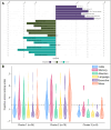
Figure 6.. Significant differences between CSF clusters and Cognitive phenotypes of CSF clusters
Panel A shows significant differences between clusters: the length of bars represents the size of significance according to p values, while the side of the bar indicates the cluster where the variable is increased/more common (first comparison C2 on the right, second and third comparison C3 on the right). Panel B shows violin plots for the Global and Domain Deficit Scores across the clusters; the dotted line cuts at 0.5, threshold to discriminate between impaired (above) and non-impaired cognitive performance (below).

References

    1. Link CD. Is There a Brain Microbiome? Neurosci Insights. 2021;16: 26331055211018709. doi:10.1177/26331055211018709 - DOI - PMC - PubMed
    1. Liang G, Bushman FD. The human virome: assembly, composition and host interactions. Nat Rev Microbiol. 2021;19: 514–527. doi:10.1038/s41579-021-00536-5 - DOI - PMC - PubMed
    1. Moustafa A, Xie C, Kirkness E, Biggs W, Wong E, Turpaz Y, et al. The blood DNA virome in 8,000 humans. PLoS Pathog. 2017;13: e1006292. doi:10.1371/journal.ppat.1006292 - DOI - PMC - PubMed
    1. Rascovan N, Duraisamy R, Desnues C. Metagenomics and the Human Virome in Asymptomatic Individuals. Annu Rev Microbiol. 2016;70: 125–141. doi:10.1146/annurev-micro-102215-095431 - DOI - PubMed
    1. Cao Z, Sugimura N, Burgermeister E, Ebert MP, Zuo T, Lan P. The gut virome: A new microbiome component in health and disease. eBioMedicine. 2022;81. doi:10.1016/j.ebiom.2022.104113 - DOI - PMC - PubMed

Publication types

LinkOut - more resources