Review
. 2025 Jan 21;4(1):102439.
doi: 10.1016/j.jscai.2024.102439. eCollection 2025 Jan.

Understanding Right Heart Flow: Implications for Interatrial Shunt Device Therapy in Heart Failure

Affiliations
Review

Understanding Right Heart Flow: Implications for Interatrial Shunt Device Therapy in Heart Failure

Raviteja Guddeti et al. J Soc Cardiovasc Angiogr Interv. .

Abstract

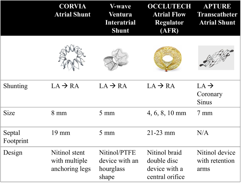
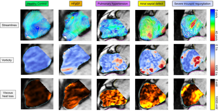
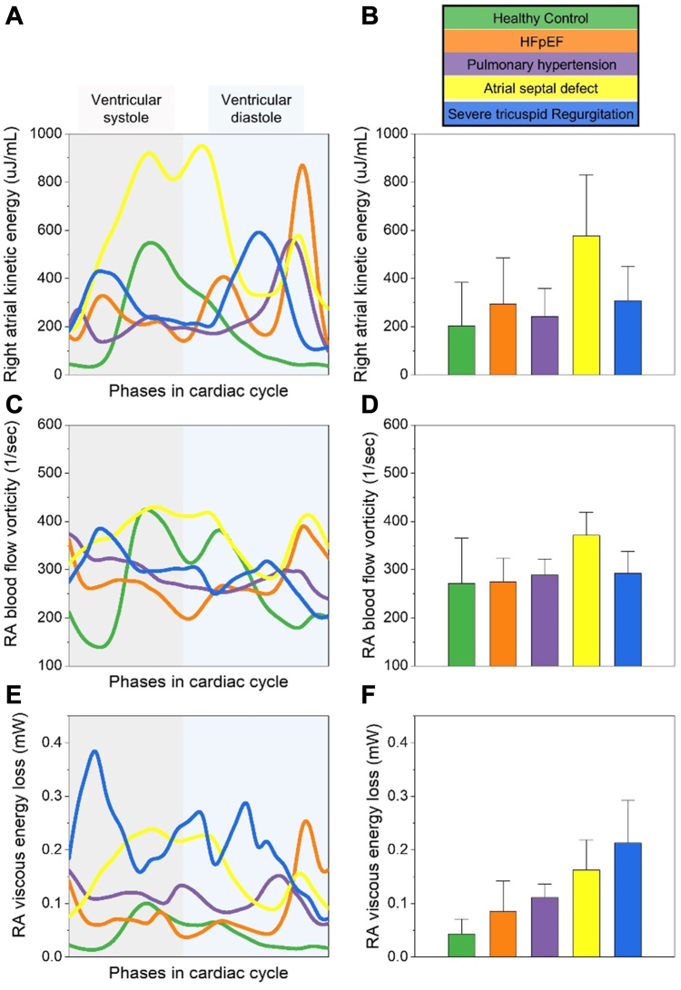
Elevation in left atrial pressure with subsequent pulmonary congestion is central to the pathology of heart failure. Interatrial shunts have emerged as a potential therapeutic strategy in patients with heart failure, especially those with diastolic dysfunction. These devices decrease left atrial pressure by shunting blood into the right atrium. Normal right heart flow is characterized by a predominant vortex formation in the right atrium, which then enters the right ventricle as a direct flow that preserves kinetic energy and right ventricular work efficiency. Examining the abnormal right heart blood flow patterns in naturally occurring interatrial shunts using 4-dimensional flow magnetic resonance imaging can improve our understanding of the effects of various interatrial shunt devices currently being investigated for heart failure management.

Keywords: heart failure; interatrial shunts; magnetic resonance imaging; right heart flow.


Figures

Figure 1
Figure 1
Normal RV flow patterns as visualized by 4D flow magnetic resonance imaging. Pathline visualization of RV blood flow during late diastolic filling in a 70-year-old healthy individual with a left ventricular end-diastolic volume index of 61 mL/m2. The inflowing blood is channeled toward the outflow tract through the ring vortex (indicated by the white arrow). Different flow components are represented as follows: direct flow (green), retained inflow (yellow), delayed ejection flow (blue), and residual volume (red). Some pathlines in the lateral region of the RV have been omitted to reveal those near the interventricular septum. A semitransparent 4-chamber image provides anatomical orientation. LA, left atrium; RA, right atrium; RV, right ventricle; RVOT, right ventricular outflow tract. Reproduced with permission from Fredriksson et al.
Figure 2
Figure 2
Partial anomalous pulmonary venous return and a sinus venosus ASD. Coronal (A) and sagittal (B) views show anomalous right upper pulmonary veins (red arrows) draining to the superior vena cava (white arrow). (C) Oblique four-chamber view shows flow from the left atrium crossing into the right atrium through the atrial septal defect (arrow). Reproduced with permission from Vasanawala et al.
Figure 3
Figure 3
Interatrial shunt devices in heart failure. AFR, atrial flow regulator; LA, left atrium; N/A, not applicable; PTFE, polytetrafluoroethylene; RA, right atrium.
Central Illustration
Visualization of intra-atrial flow patterns, vorticity, and viscous heat loss in various cardiac conditions. Intra-atrial hemodynamics for a healthy individual (control) and cases of HFpEF, pulmonary hypertension, atrial septal defect, and severe tricuspid regurgitation are shown during atrial filling coinciding with ventricular systole. Rows depict streamlines (top, 0-40 cm/s), vorticity (middle, 0-1000 1/s), and viscous heat loss (bottom, 0-10,000 mW). Healthy individual shows orderly flow, minimal vorticity, and low heat loss. The organized vortical flow appears to reduce heat loss and effectively reduce the overall vorticity. In the HFpEF case, the central organized vortical flow seen in the control is missing, and there is a bit of disorganized flow in streamline visualization, which possibly results in a minor increase in vorticity and heat loss. In a patient with pulmonary hypertension, there is a significant increase in disorganized streamlines in the right atrium. This complements the associated increase in vorticity and heat loss observed. In a case of atrial septal defect, there is a very high velocity signal across the septum in streamline visualization, which appears to result in high vorticity and heat loss. Similar observations are seen in a patient with severe tricuspid regurgitation.
Figure 4
Figure 4
RA blood flow kinetic energy, vorticity, and viscous energy loss in various cardiac conditions. (A) RA kinetic energy (μJ/mL) during cardiac cycle phases. (B) Mean RA kinetic energy with SD. (C) RA blood flow vorticity (1/s) over the cardiac cycle phases. (D) Mean RA blood flow vorticity with SD. (E) RA viscous energy loss (mW) during cardiac cycle phases. (F) Mean RA viscous energy loss with SD. Each condition is color-coded: healthy control (green), severe tricuspid regurgitation (orange), HFpEF with AF (purple), pulmonary hypertension (yellow), and atrial septal defect (blue). AF, atrial fibrillation; HFpEF, heart failure with preserved ejection fraction; RA, right atrial.

References

    1. Pfeffer M.A., Shah A.M., Borlaug B.A. Heart failure with preserved ejection fraction in perspective. Circ Res. 2019;124(11):1598–1617. - PMC - PubMed
    1. Shah S.J., Borlaug B.A., Chung E.S., et al. Atrial shunt device for heart failure with preserved and mildly reduced ejection fraction (REDUCE LAP-HF II): a randomised, multicentre, blinded, sham-controlled trial. Lancet. 2022;399(10330):1130–1140. - PubMed
    1. Haddad F., Hunt S.A., Rosenthal D.N., Murphy D.J. Right ventricular function in cardiovascular disease, part I: anatomy, physiology, aging, and functional assessment of the right ventricle. Circulation. 2008;117(11):1436–1448. - PubMed
    1. Bolger A.F., Heiberg E., Karlsson M., et al. Transit of blood flow through the human left ventricle mapped by cardiovascular magnetic resonance. J Cardiovasc Magn Reson. 2007;9(5):741–747. - PubMed
    1. Lahm T., Douglas I.S., Archer S.L., et al. Assessment of right ventricular function in the research setting: knowledge gaps and pathways forward. An official American Thoracic Society research statement. Am J Respir Crit Care Med. 2018;198(4):e15–e43. - PMC - PubMed

LinkOut - more resources