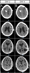
An Unusual Presentation of Peri-Lead Edema Following Deep Brain Stimulation for Parkinson's Disease: A Case Report and Review of the Literature
- PMID: 40062311
- PMCID: PMC11885058
- DOI: 10.1002/ccr3.70221
An Unusual Presentation of Peri-Lead Edema Following Deep Brain Stimulation for Parkinson's Disease: A Case Report and Review of the Literature
Abstract
Peri-lead edema (PLE) after deep brain stimulation may mimic brain infection on magnetic resonance imaging (MRI). We present a case of symptomatic PLE with annular contrast enhancement on MRI suggestive of an infectious cause. We show that careful clinical evaluation and laboratory testing, in addition to neuroimaging, are essential to guide treatment and to avoid unnecessary interventions in PLE cases with a favorable spontaneous course.
Keywords: Parkinson's disease; deep brain stimulation; movement disorders; neuromodulation; peri‐lead edema.
© 2025 The Author(s). Clinical Case Reports published by John Wiley & Sons Ltd.
Conflict of interest statement
M.W. is working as a consultant for Stryker Neurovascular. M.W. has been working as a consultant for Kaneka Pharmaceuticals and Medtronic (both inactive). M.W. has received reimbursement for lectures or travel support from Bracco Imaging, Medtronic, Siemens Healthcare, and Stryker Neurovascular. M.W. has received grants for research projects or educational exhibits from ab medica, Acandis, Bayer, Bracco Imaging, Cerenovus, Codman Neurovascular, Dahlhausen, Kaneka Pharmaceuticals, Medtronic, Mentice AB, Microvention, Phenox, Siemens Healthcare, and Stryker Neurovascular. J.B.S. is working in the advisory board of Forward Pharma, MSD, Lundbeck, Biogen, Eisai, Novo Nordisk, Roche, Reata, and Lilly. J.B.S. has received reimbursement for lectures from Merz, Teva, Bayer, UCB, Lilly, Boehringer, GSK, Bial, Novartis, Biogen, and Eisai. J.B.S. has received grants for research projects from Biogen, Eisai, and Lilly. F.H. received travel and conference fees from Bial, Desitin, Abbott, Zambon, and Abbvie. V.S.W. received travel and conference fees from Medtronic. The other authors declare no conflicts of interest.
Figures


Similar articles
-
Decreased brain volume may be associated with the occurrence of peri-lead edema in Parkinson's disease patients with deep brain stimulation.Parkinsonism Relat Disord. 2024 Apr;121:106030. doi: 10.1016/j.parkreldis.2024.106030. Epub 2024 Feb 9. Parkinsonism Relat Disord. 2024. PMID: 38354427
-
[Early brain imaging changes and its influence on electrode impedance after implantation of 3.0 T MRI-compatible deep brain stimulation system in Parkinson's disease subthalamic nucleus].Zhonghua Yi Xue Za Zhi. 2023 Dec 19;103(47):3809-3815. doi: 10.3760/cma.j.cn112137-20231009-00682. Zhonghua Yi Xue Za Zhi. 2023. PMID: 38123221 Chinese.
-
Acute symptomatic peri-lead edema 33 hours after deep brain stimulation surgery: a case report.J Med Case Rep. 2017 Apr 14;11(1):103. doi: 10.1186/s13256-017-1275-6. J Med Case Rep. 2017. PMID: 28407815 Free PMC article.
-
The need to be alert to complications of peri-lead cerebral edema caused by deep brain stimulation implantation: A systematic literature review and meta-analysis study.CNS Neurosci Ther. 2022 Mar;28(3):332-342. doi: 10.1111/cns.13802. Epub 2022 Jan 19. CNS Neurosci Ther. 2022. PMID: 35044099 Free PMC article.
-
Deep brain stimulation and genetic variability in Parkinson's disease: a review of the literature.NPJ Parkinsons Dis. 2019 Sep 6;5:18. doi: 10.1038/s41531-019-0091-7. eCollection 2019. NPJ Parkinsons Dis. 2019. PMID: 31508488 Free PMC article. Review.
Cited by
-
Unveiling patterns of peri-lead edema after deep brain stimulation: a retrospective review of clinical and demographic factors.Neuroradiology. 2025 Apr 8. doi: 10.1007/s00234-025-03607-z. Online ahead of print. Neuroradiology. 2025. PMID: 40198366
References
-
- Haberler C., Alesch F., Mazal P. R., et al., “No Tissue Damage by Chronic Deep Brain Stimulation in Parkinson's Disease,” Annals of Neurology 48, no. 3 (2000): 372–376. - PubMed
LinkOut - more resources
Full Text Sources

