. 2025 Oct 1;24(10):1626-1639.
doi: 10.1158/1535-7163.MCT-24-0906.

Specific Genetic Mutations Impact Chemotherapy Resistance and Therapeutic Efficacy of Oncolytic Viruses in Ovarian Cancer

Affiliations

Specific Genetic Mutations Impact Chemotherapy Resistance and Therapeutic Efficacy of Oncolytic Viruses in Ovarian Cancer

Alison O Cudmore et al. Mol Cancer Ther. .

Abstract

Epithelial ovarian cancer (EOC) is the most lethal gynecologic cancer, and those affected are in urgent need of new therapeutic strategies. Standard treatment is surgery followed by taxane- and platinum-based chemotherapy. However, the rate of relapse is high, and the 5-year survival is only 45%. Oncolytic viruses (OV) are a promising approach to EOC therapy through remodeling the immune composition of the tumor microenvironment. Treatment response in EOC tumors can differ based on the presence of key tumorigenic mutations. This study evaluated the impact of specific tumor mutations on the response to the current standard-of-care carboplatin, two promising OV candidates VSVΔM51 and MG1, an infected cell vaccine (ICV-MG1) regimen, and the antiangiogenic drug Fc3TSR. Mice with tumors harboring constitutive K-Ras activation showed an enhanced response to carboplatin and VSVΔM51 treatment. Additionally, VSVΔM51 treatment prolonged survival of syngeneic mice bearing tumors with mutations in Pten and Kras, Pten and Trp53, or Trp53 and Brca2 with increased activation of CD4+ and CD8+ T lymphocytes in the peritoneal tumor microenvironment. To enhance OV potency, an MG1-based infected cell vaccine inducing the expression of IL21 or IL15 + IL21 was developed and found to enable strong and long-lasting antitumoral immunity in two carboplatin-refractory syngeneic models, ID8-Trp53-/- and STOSE. VSVΔM51 combined with the antiangiogenic Fc3TSR enhanced efficacy in the ID8 model. In summary, OV-based immunotherapy has shown promise in diverse murine models of EOC-bearing clinically relevant mutations, thus laying the foundation for developing new OV-based strategies to target a large spectrum of EOC genotypes.


Conflict of interest statement

A.O. Cudmore reports grants from Ovarian Cancer Canada and Canadian Institutes of Health Research during the conduct of the study. H. Murshed reports grants from Ottawa Hospital Research Institute during the conduct of the study. E.A. Macdonald reports grants from Canadian Institutes of Health Research and Ovarian Cancer Canada during the conduct of the study. M. Grondin reports grants from Canadian Institutes of Health Research and Ovarian Cancer Canada during the conduct of the study. B.C. Vanderhyden reports grants from Canadian Institutes of Health Research and Ovarian Cancer Canada during the conduct of the study. No disclosures were reported by the other authors.

Figures

Figure 1.
Figure 1.
Sensitivity of murine HGSC models to a high-dose regimen of carboplatin treatment. A, Absolute IC50 doses of carboplatin on in vitro murine models of ovarian cancer. Results were analyzed by one-way ANOVA with the Tukey post hoc test (n = 3). B, Survival benefit of carboplatin treatment in tumor-bearing mice in vivo. After tumor establishment, mice were treated with a biweekly injection of carboplatin at 20 mg/kg per mouse for a total of 4 weeks (8 doses). Results were analyzed by the log-rank Mantle–Cox test. The median survival is indicated in the parentheses in the figure (n = 8–10 mice per treatment group). (*, P < 0.05; **, P < 0.01; ***, P < 0.001; ****, P ≤ 0.0001).
Figure 2.
Figure 2.
Screening of two OV platforms in vitro against murine ovarian cancer cell lines. A and B, alamarBlue assay of cells infected with (A) Maraba MG1 and (B) VSVΔM51, at different MOIs for the lengths of time indicated on the y-axis (n = 3). Data were analyzed by two-way ANOVA followed by the Tukey post hoc test. *, P < 0.05; **, P < 0.01; ****, P < 0.0001. C and D, Induction of IFN-β in murine cell lines 6 and 12 hours after VSVΔM51 infection relative to 0-hour control. Data were normalized to the level of expression in untreated cells (n = 3).
Figure 3.
Figure 3.
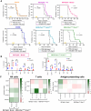
VSVΔM51 treatment confers antitumoral protection against ovarian cancer in syngeneic models. C57BL/6 mice bearing intraperitoneal tumors were treated starting at 25% of expected length of survival with 3 × 10⁸ PFU/mouse of VSVΔM51 by intraperitoneal injection every 3 days, for a total of three doses or PBS control. A, Kaplan–Meier plot of survival of VSVΔM51-treated tumor-bearing mice. Results were analyzed by the log-rank Mantle–Cox test. B, Immune cell populations found in the peritoneal cavity 5 days after the last treatment of tumor-bearing mice. The total population of leukocytes is defined as CD45+ live cells (See Supplementary Fig. S2). Analysis by one-way ANOVA with the Dunnett post hoc test. *, P < 0.05; **, P < 0.01; ****, P < 0.0001. NK-T, Natural killer T cells; TAM, tumor-associated macrophages. C, Fold induction relative to PBS treatment of activation markers on NK cells (left), T lymphocytes (middle), and antigen-presenting cells (right) after treatment in peritoneal wash samples of tumor-bearing mice.
Figure 4.
Figure 4.
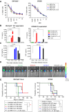
Delivery of IL15 and IL21 with an ICV confers significant antitumoral protection. A, alamarBlue was used to determine cell viability of ID8-Trp53−/− and STOSE cell lines 24 hours after infection with MG1 variants at the indicated MOIs. B, IL15 and (C) IL21 secretion were assessed by ELISA of supernatants from infected cells (ICV) after 2 and 24 hours (n = 3); mean ± SD. D and E, Mice were injected with STOSE or ID8-Trp53−/−GLuc cells and treated at day 11 with 2.5 × 10⁷ ICV-MG1 variants. To monitor tumor burden, (D) IVIS imaging was performed at day 47 after cell injection. Irr cells, irradiated cells. E, Kaplan–Meier plot of survival was analyzed by the log-rank Mantle–Cox test (n = 4–6 mice/group). *, P < 0.05; **, P < 0.01; ***, P < 0.001; ****, P ≤ 0.0001.
Figure 5.
Figure 5.
Three doses of ICV delivering IL15 and IL21 confer superior protection against ovarian tumor growth. Eight-week-old mice were injected with (A) ID8-Trp53−/−GLuc or (B) STOSE cells and treated on days 7, 10, and 13 with 2.5 × 10⁷ ICV-MG1 variants (see Supplementary Fig. S1A). Survival shown by the Kaplan–Meier plot analyzed by the log-rank Mantle–Cox test. n = 5–7 mice/group. C and E, Tumor-naïve mice received one intraperitoneal dose of 2.5 × 10⁷ ICV-MG1 variants, and peritoneal washes were collected 24 hours later to study immune cell recruitment into the TME. C, Total leukocytes, DX5+ NK cells, and CD4/CD8 T-cell ratio in the peritoneal TME. D, MHC-II and PD-L1 expression on CD11c+ DC1 and CD11b+F4/80+ macrophages in the peritoneal TME. E, PD-1 expression on DX5+ NK and CD8+ T cells in the peritoneal TME. Gating strategy shown in Supplementary Fig. S2. n = 4 mice/group. *, P < 0.05; **, P < 0.01; ***, P < 0.001; ****, P< 0.0001.
Figure 6.
Figure 6.
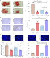
The antiangiogenic compound Fc3TSR and VSV induce tumor regression and vascular normalization in an orthotopic, syngeneic mouse model of ovarian cancer. After 50 days of tumor growth, mice were injected with PBS, Fc3TSR, or VSV alone or in combination. A, Tumor weights following treatment. B, Incidence of apoptotic cell death. Scale bar, 50 µm. C, Tissues were co-stained to visualize endothelial (CD31, red) and smooth muscle (α-SMA, green) cells to provide an index of vessel maturity. Analysis by one-way ANOVA; *, P < 0.05; **, P < 0.01; ****, P< 0.0001.

References

    1. Baldwin LA, Huang B, Miller RW, Tucker T, Goodrich ST, Podzielinski I, et al. Ten-year relative survival for epithelial ovarian cancer. Obstet Gynecol 2012;120:612–8. - PubMed
    1. Huang J, Chan WC, Ngai CH, Lok V, Zhang L, Lucero-Prisno DE, et al. Worldwide burden, risk factors, and temporal trends of ovarian cancer: a global study. Cancers 2022;14:2230. - PMC - PubMed
    1. Lheureux S, Gourley C, Vergote I, Oza AM. Epithelial ovarian cancer. The Lancet 2019;393:1240–53. - PubMed
    1. Eddie SL, Quartuccio SM, Ó hAinmhir E, Moyle-Heyrman G, Lantvit DD, Wei J-J, et al. Tumorigenesis and peritoneal colonization from fallopian tube epithelium. Oncotarget 2015;6:20500–12. - PMC - PubMed
    1. Quartuccio SM, Karthikeyan S, Eddie SL, Lantvit DD, Ó hAinmhire E, Modi DA, et al. Mutant p53 expression in fallopian tube epithelium drives cell migration. Int J Cancer 2015;137:1528–38. - PMC - PubMed

MeSH terms