. 2025 Apr 1;328(4):G433-G447.
doi: 10.1152/ajpgi.00337.2024. Epub 2025 Mar 10.

Cinnabarinic acid protects against metabolic dysfunction-associated steatohepatitis by activating aryl hydrocarbon receptor-dependent AMPK signaling

Affiliations

Cinnabarinic acid protects against metabolic dysfunction-associated steatohepatitis by activating aryl hydrocarbon receptor-dependent AMPK signaling

Nikhil Y Patil et al. Am J Physiol Gastrointest Liver Physiol. .

Abstract

Metabolic dysfunction-associated steatohepatitis (MASH) is an advanced form of metabolic dysfunction-associated steatotic liver disease (MASLD) characterized by the accumulation of fats in the liver, chronic inflammation, hepatocytic ballooning, and fibrosis. This study investigates the significance of hepatic aryl hydrocarbon receptor (AhR) signaling in cinnabarinic acid (CA)-mediated protection against MASH. Here, we report that livers of high-fat, high-fructose, high-cholesterol diet-fed hepatocyte-specific aryl hydrocarbon receptor knockout mice (AhR-hKO) exhibited aggravated steatosis, inflammation, and fibrosis compared with control AhR-floxed livers. Moreover, treatment with a tryptophan catabolite, CA, reduced body weight gain and significantly attenuated hepatic steatosis, inflammation, ballooning, fibrosis, and liver injury only in AhR-floxed but not in AhR-hKO mice, strongly indicating that the CA-mediated protection against steatohepatitis is AhR-dependent. Furthermore, protection against lipotoxicity by CA-activated AhR signaling was confirmed by utilizing an in vitro human hepatocyte model of MASLD. Mechanistically, CA-induced AhR-dependent signaling augmented AMP-activated protein kinase (AMPK), leading to the upregulation of peroxisome proliferator-activated receptor gamma coactivator-1alpha (PGC1α) and attenuation of sterol regulatory element-binding protein-1 (SREBP1) to regulate hepatic lipid metabolism. Collectively, our findings indicate that CA-mediated protection against MASH is dependent on hepatic AhR signaling, and selective endogenous AhR agonists that regulate lipogenesis can serve as promising future therapeutics against MASLD.NEW & NOTEWORTHY The study showed that the absence of AhR in hepatocytes results in exacerbated metabolic dysfunction-associated steatohepatitis (MASH) in mice subjected to a Western-style high-fat, high-fructose, high-cholesterol diet. Moreover, treatment with a tryptophan catabolite, cinnabarinic acid (CA), mitigated hallmarks of MASH in an AhR-dependent manner. In conclusion, the study delineates the significance of hepatic AhR-dependent AMPK signaling in CA-mediated protection against MASH.

Keywords: aryl hydrocarbon receptor; cinnabarinic acid; liver; metabolic dysfunction-associated steatohepatitis; metabolic dysfunction-associated steatotic liver disease.


Conflict of interest statement

DISCLOSURES

The authors declare no conflict of interest.

Figures
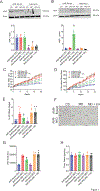
Fig. 1
Fig. 1. CA-mediated attenuation of body mass and adiposity is dependent on hepatic AhR.
AhR-floxed (AhRfl/fl, control) and hepatocyte-specific AhR knockout (AhR-hKO) mice were fed a control (CD) or a high-fat, high-fructose, high-cholesterol diet (MASH diet, MD) for 20 weeks. 12 mg/kg CA was intraperitoneally injected to AhR-floxed and AhR-hKO mice for 20 weeks (MD + CA). After 20 weeks, western blotting was performed to detect AhR expression. Representative western blot of AhR in A) whole-liver lysates, normalized to actin as a loading control and to AhR-floxed (CD) group, B) liver nuclear extracts (normalized to lamin control and to AhR-floxed (CD) cohort), C) body mass, D) normalized body mass, E) % gonadal white adipose tissue to body mass at the end of the study, week 20, F) representative H&E staining of white adipose tissue, scale bar = 200 μm, G) Average white adipocyte area calculated with ImageJ software from 68–112 cells/mouse, H) average daily food intake calculated from average weekly food intake. Data are represented as mean ± SD (n=7). *p<0.05 compared to AhR-floxed (CD) group for A-B, and to the AhR-floxed (MD) group for C-H.
Fig. 2
Fig. 2. CA treatment improves MASH pathology only in AhR-floxed mice.
A) Representative image of AhR-floxed and AhR-hKO mice liver after 20 weeks of control diet, MASH diet, and MASH diet + CA treatment. B) percentage ratio of liver weight to body weight, C) representative H&E image of liver sections (scale bar = 200 μm), S = steatosis, and B = Ballooning in zoomed images, D) representative Oil Red O staining of liver sections (scale bar = 200 μm), E) quantitation of oil red O positive area shown as a percentage of total area, F) liver triglyceride, G) liver cholesterol, H) relative mole percentage of predominant free fatty acid species in livers of MD and MD + CA administered mice, I) total hepatic free fatty acids (nmole/mg tissue) of liver, J) picrosirius red staining of liver sections, scale bar = 200 μm K) quantitation of sirius red positive area using ImageJ (4 to 6 fields/mice), L) mRNA expression of fibrosis markers analyzed by qRT-PCR and normalized to 18S ribosomal RNA, M) liver F4/80 staining (scale bar = 200 μm), N) mRNA expression of inflammatory genes in liver tissue normalized to 18S rRNA, O) serum ALT measurement as an indicator of liver injury, P) NAS and Brunt fibrosis score. All results are shown as mean ± SD. Univariate ANOVA and Dunnett’s post hoc were used to calculate the p values. * P <0.05 compared with AhR-floxed (MD). n = 7 mice per group.
Fig. 3
Fig. 3. CA improves glucose tolerance in AhR-floxed mice.
A) fasting blood glucose measurements upon completion of 20 weeks of control diet, MASH diet, and MASH diet + CA treatments, B) glucose tolerance test, C) the area under the curve (AUC) was calculated for all six experimental cohorts. Data represented as mean ± SD (n = 7). *p<0.05 compared to AhR-floxed MD-only treatment group.
Fig. 4
Fig. 4. Liver transcriptomic analysis revealed enrichment of AMPK signaling with CA treatment in AhR-floxed mice.
Liver RNA isolated from AhR-floxed CD, MD, MD + CA groups (four randomly selected mice from each group) was used for RNA sequencing. A) Volcano plot showing comparison of differentially expressed genes B) heatmap indicating top 100 differentially expressed genes, C) heatmap illustrating Hallmark gene expression sets ranked by lowest ANOVA p values across three groups (CD, MD, MD + CA), D) Enriched kinase pathways were determined from genes upregulated in MD + CA group compared to MD-only cohort by using ARCHS4 Kinase Library through the web-based analysis tool Enrichr, E) heatmap showing the expression of genes involved in AMPK signaling between CD, MD, MD + CA group from Hallmark library, F) heatmap visualizing AMPK signaling pathway scores, G) ssGSEA enrichment score for AMPK signaling. Data represented as mean ± SD (n = 4). *p<0.05 compared to MD.
Fig. 5
Fig. 5. CA-induced AhR protects against lipotoxicity by activating AMPK signaling.
A) Representative western blot for phospho-AMPK and AMPK from livers of AhR-floxed and AhR-hKO mice (CD, MD, MD + CA groups), B) expression of p-AMPK normalized to AMPK and compared to AhR-floxed (CD), C) immunofluorescence was performed on AhR-floxed and AhR-hKO liver tissue sections probing for pAMPK (green) (scale bar = 200 μm), D) AhR-floxed and AhR-hKO mouse primary hepatocytes were isolated and treated with vehicle (DMSO), CA (30 μm), OA (500 μM), and OA + CA simultaneously for 24 hr. For compound C treatment, 20 μM compound C was added to AhR-floxed mouse hepatocytes 2 hours prior to the CA/OA treatments. Representative western blots of p-AMPK and AMPK are shown. E) expression of p-AMPK/AMPK compared to AhR-floxed (V) in mouse primary hepatocytes, F) representative images of Oil Red O staining (scale bar = 200 μm), G) quantitation of Oil Red O positive area calculated by ImageJ (2 to 3 fields/mice), H) representative western blotting for PGC1α from whole liver lysates of AhR-floxed and AhR-hKO mice, I) expression of PGC1α normalized to actin and AhR-floxed (CD), J) immunohistochemistry to detect expression of PGC1α in AhR-floxed and AhR-hKO liver sections (red) (scale bar = 200 μm), K) mRNA message of PGC1α normalized to 18S rRNA from liver, L) mRNA expression of markers for de novo lipogenesis and triglyceride synthesis analyzed by qRT-PCR and normalized to 18S rRNA from liver, M) representative western blot for SREBP1, N) protein expression of SREBP1 normalized to actin and AhR-floxed (CD). Data represented as mean ± SD (n = 7 for in vivo studies and n = 3 or 4 for isolated mouse primary hepatocytes). *p<0.05 compared to AhR-floxed MD-only treatment group for in vivo studies and to AhR-floxed OA treated cohort for in vitro studies.
Fig. 6
Fig. 6. CA-induced AhR-AMPK activation protects human hepatocytes against lipotoxicity.
A) Cryopreserved human hepatocytes were treated with vehicle, oleic acid (OA, 200 μM), and simultaneously with oleic acid, 200 μM OA and cinnabarinic acid, 30 μM, 50 μM, 100 μM CA for 24 hours. Accumulation of lipid droplets was visualized using BODIPY 493/503 staining. Human hepatocytes were transiently transfected with non-targeting (NT siRNA) or AhR siRNA for 24 hours followed by vehicle (DMSO), 200 μM OA, and 200 μM OA and 30 μM CA treatment concomitantly for another 24 hr. B) western blotting on total human hepatocyte lysates was performed to detect AhR expression, C) quantitation of AhR expression normalized to actin and compared to NT siRNA (V), D) BODIPY staining to localize neutral lipid droplets in non-targeting and AhR siRNA-transfected vehicle, OA, and OA+CA-treated human hepatocytes, E) western blotting to monitor p-AMPK, AMPK, PGC1α, and SREBP1 expression. Actin was used to normalize PGC1α, and SREBP1 expression, F) quantitation of p-AMPK/AMPK normalized to NT siRNA (V), G) quantitation of PGC1α normalized to actin and to NT siRNA (V), H) quantitation of SREBP1 from total human hepatocyte lysates normalized to actin and to NT siRNA (V). Data are represented as mean ± SD (n=3). *p<0.05 compared to NT siRNA (OA).

References

    1. Wang X, Zhang L, and Dong B. Molecular mechanisms in MASLD/MASH-related HCC. Hepatology 1–22, 2024. - PMC - PubMed
    1. Shetty A, and Syn WK. Health and Economic Burden of Nonalcoholic Fatty Liver Disease in the United States and Its Impact on Veterans. Fed Pract 36: 14–19, 2019. - PMC - PubMed
    1. Harrison SA, Bedossa P, Guy CD, Schattenberg JM, Loomba R, Taub R, Labriola D, Moussa SE, Neff GW, Rinella ME, Anstee QM, Abdelmalek MF, Younossi Z, Baum SJ, Francque S, Charlton MR, Newsome PN, Lanthier N, Schiefke I, Mangia A, Pericas JM, Patil R, Sanyal AJ, Noureddin M, Bansal MB, Alkhouri N, Castera L, Rudraraju M, Ratziu V, and Investigators M-N. A Phase 3, Randomized, Controlled Trial of Resmetirom in NASH with Liver Fibrosis. The New England journal of medicine 390: 497–509, 2024. - PubMed
    1. Lowe MM, Mold JE, Kanwar B, Huang Y, Louie A, Pollastri MP, Wang C, Patel G, Franks DG, Schlezinger J, Sherr DH, Silverstone AE, Hahn ME, and McCune JM. Identification of cinnabarinic acid as a novel endogenous aryl hydrocarbon receptor ligand that drives IL-22 production. PloS one 9: e87877, 2014. - PMC - PubMed
    1. Rao PV, and Vaidyanahan CS. Enzymic conversion of 3-hydroxyanthranilic acid into cinnabarinic acid. Partial purification and properties of rat-liver cinnabarinate synthase. The Biochemical journal 99: 317–322, 1966. - PMC - PubMed

MeSH terms

Substances

LinkOut - more resources