. 2025 Apr;13(4):e0269224.
doi: 10.1128/spectrum.02692-24. Epub 2025 Mar 10.

Application of versatile reverse genetics system for feline coronavirus

Affiliations

Application of versatile reverse genetics system for feline coronavirus

Izumi Kida et al. Microbiol Spectr. 2025 Apr.

Abstract

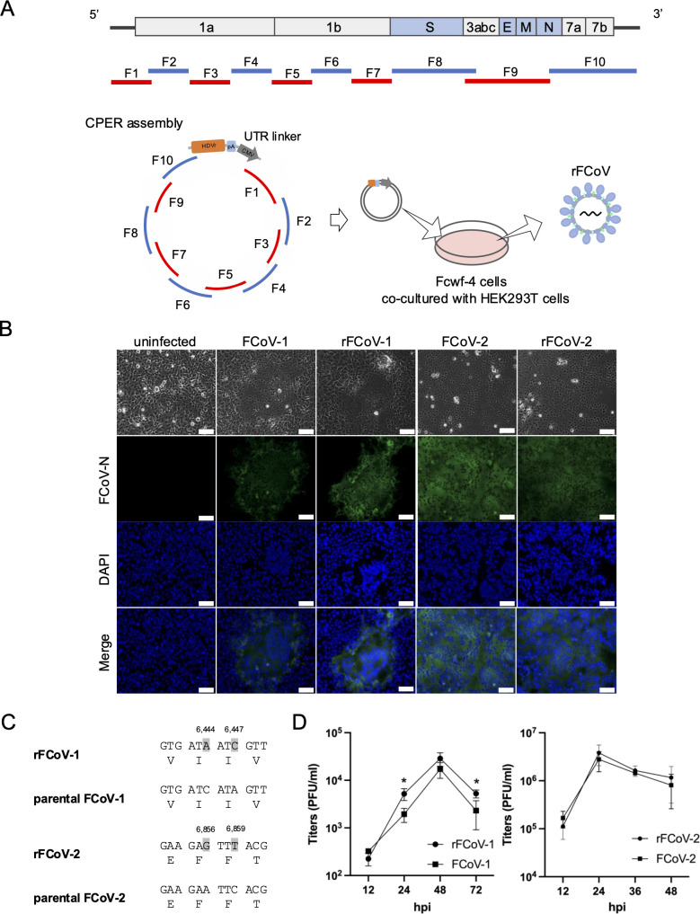
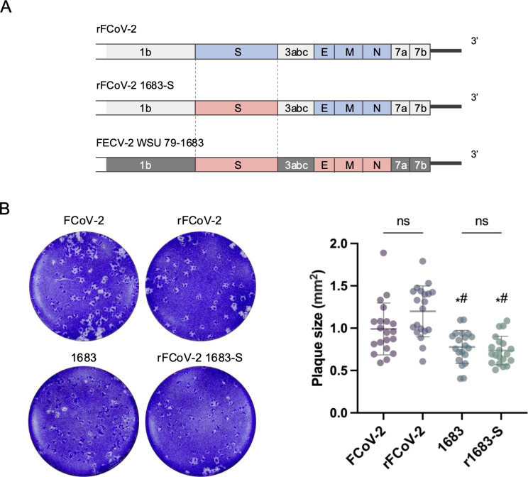
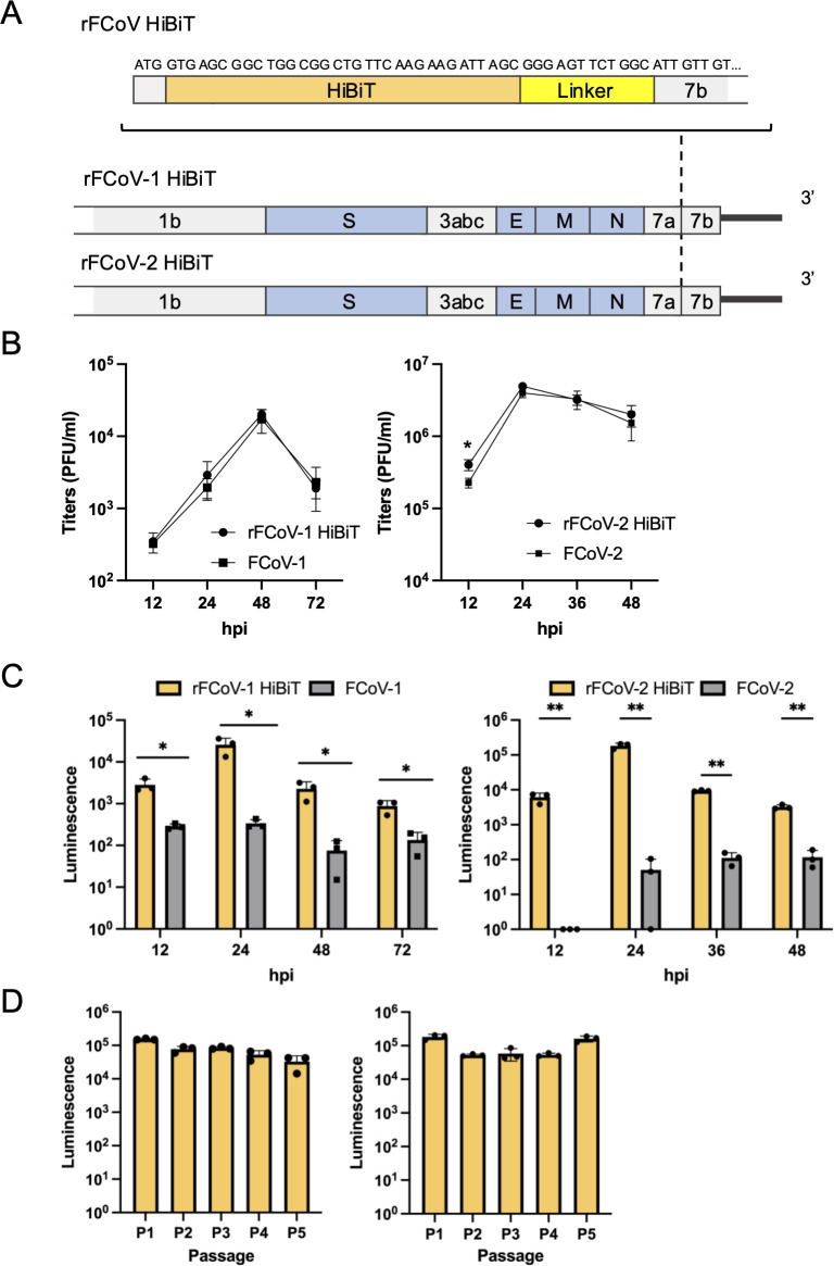
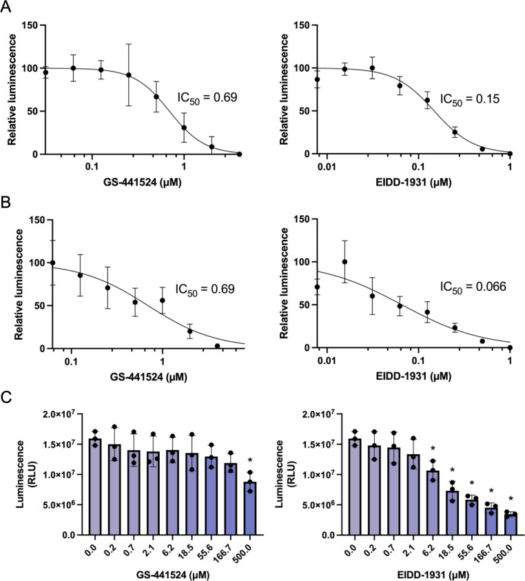
Feline infectious peritonitis (FIP) is a fatal disease caused by feline coronavirus (FCoV). Although multiple gene mutations in FCoV likely account for FIP pathogenesis, molecular studies for FCoV have been limited due to the lack of a suitable reverse genetics system. In the present study, we established a rapid PCR-based system to generate recombinant FCoV using the circular polymerase extension reaction (CPER) method for both serotype 1 and 2 viruses. Recombinant FCoV was successfully rescued at sufficient titers to propagate the progeny viruses with high sequence accuracy. The growth kinetics of recombinant FCoV were comparable to those of the parental viruses. We successfully generated recombinants harboring the spike gene from a different FCoV strain or a reporter HiBiT tag using the CPER method. The chimeric virus demonstrated similar characteristics with the parental virus of the spike gene. The reporter tag stably expressed after five serial passages in the susceptible cells, and the reporter virus could be applied to evaluate the sensitivity of antiviral inhibitors using the luciferase assay system to detect HiBiT tag. Taken together, our versatile reverse genetics system for FCoV shown herein is a robust tool to characterize viral genes even without virus isolation and to investigate the molecular mechanisms of the proliferation and pathogenicity of FCoV.

Importance: Feline infectious peritonitis is a highly fatal disease in cats caused by feline coronavirus variants that can infect systemically. Due to the lack of a versatile toolbox for manipulating the feline coronavirus genome, an efficient method is urgently needed to study the virus proteins responsible for the severe disease. Herein, we established a rapid reverse genetics system for the virus and demonstrated the capability of the recombinant viruses to be introduced with desired modifications or reporter genes without any negative impacts on virus characteristics in cell culture. Recombinant viruses are also useful to evaluate antiviral efficacy. Overall, our system can be a promising tool to reveal the molecular mechanisms of the viral life cycle of feline coronavirus and disease progression of feline infectious peritonitis.

Keywords: feline coronavirus; feline infectious peritonitis; reverse genetics.


Conflict of interest statement

The authors declare no conflict of interest.

Figures

Fig 1
Fig 1
Establishment of reverse genetics for FCoV by CPER. (A) Schematic representation of recombinant virus rescue. FCoV genome (top) was divided into 10 cDNA fragments (F1–F10) covering the full-length of the FCoV genome with 13 to 236 nt overlapping ends (middle) and used for a CPER assembly for generating recombinant virus (rFCoV) (bottom). The F1 to F10 fragments were assembled with a UTR linker fragment by CPER, and then the resulting CPER products were transfected into Fcwf-4 cells co-cultured with HEK293T cells. (B) Immunofluorescence assay of Fcwf-4 cells infected with parental FCoVs and CPER-generated rFCoVs. Viral antigen was visualized by staining with anti-FCoV N protein monoclonal antibody (green). Nuclei were stained with 4´,6-diamidino-2-phenylindole (DAPI) (blue); scale bar: 100 µm. (C) Genetic markers, two silent mutations (i.e., C6444A and A6447C for FCoV-1 and A6856G and C6859T for FCoV-2) were introduced into the plasmids for the corresponding fragments and confirmed in the recombinant FCoV genomes. (D) Growth kinetics of rFCoVs and parental FCoVs. Fcwf-4 cells were infected with the viruses at a multiplicity of infection of 0.01, and the virus titers in the supernatants were measured from 12 to 48 or 72 h post-infection (hpi). The presented data were expressed as mean ± SD of triplicate samples. *: P < 0.05 by two-tailed Student’s t-test without adjustment for multiple comparisons.
Fig 2
Fig 2
Construction of chimeric FCoV encoding spike gene from FECV-2. (A) Gene structures of parental viruses and chimeric FCoV encoding S gene from FECV-2. rFCoV-2 was used as a backbone for generating a chimeric FCoV expressing FECV S protein (1683-S) derived from FCoV-2 WSU 79-1683 strain, which is a serotype-2 FECV. (B) Plaque formation of parental FCoVs and rFCoVs in Fcwf-4 cells. Plaque assay was performed using Fcwf-4 cells inoculated with the parental FCoV-2, rFCoV-2, parental FECV (1683), and rFCoV-2 carrying 1683-S (r1683-S). Representative figures (left) and the sizes of plaques (n = 20 for each virus, right) are shown. Each dot in the graph indicates the diameter of a plaque. The mean ± SD was shown for each virus. Statistically significant differences versus parental FCoV-2 (*: P  <  0.05) and recombinant FCoV-2 (#: P  <  0.05) were determined by one-way analysis of variance with Tukey’s test; ns, not significant.
Fig 3
Fig 3
Construction of FCoV recombinants carrying a HiBiT gene. (A) Gene structure of FCoV recombinants carrying the HiBiT gene (rFCoV HiBiT). HiBiT gene was inserted immediately after the start codon (ATG) of the ORF7b followed by a linker. (B) Growth kinetics of rFCoVs HiBiT and parental FCoVs. Fcwf-4 cells were infected with the viruses at an MOI of 0.01, and the virus titers in the supernatants were measured from 12 to 48 or 72 h post-infection (hpi). (C) Luciferase activities in Fcwf-4 cells infected with rFCoVs HiBiT and parental FCoVs. Cells were lysed followed by the addition of LgBiT, and the luciferase activities were determined from 12 to 48 or 72 hpi. (D) Stable expression of HiBiT tag during passage recombinant viruses. rFCoVs HiBiT were passaged five times (P1–P5) in Fcwf-4 cells. Luciferase activities in cells during each passage were determined at 24 hpi. In (B–D), the presented data were expressed as mean ± SD of triplicate samples. In (B) and (C), *P < 0.05 and **P < 0.01 by a two-tailed Student’s t-test without adjustment for multiple comparisons.
Fig 4
Fig 4
Application of reporter FCoVs for assessment of antiviral inhibitors. (A) IC50 of antiviral inhibitors against recombinant FCoV-1 carrying the HiBiT gene (rFCoV-1 HiBiT). Fcwf-4 cells infected with rFCoV-1 HiBiT at an MOI of 0.01 were treated with GS-441524 and EIDD-1931 at the time of infection. The luciferase activity levels were determined by a luciferase assay at 24 h. Relative luminescence was calculated by the intensity of luciferase activity in the cells treated with antiviral inhibitors in comparison to mock control. (B) IC50 of antiviral inhibitors against recombinant FCoV-2 carrying the HiBiT gene (rFCoV-2 HiBiT). Fcwf-4 cells infected with rFCoV-2 HiBiT at an MOI of 0.001 were treated with GS-441524 and EIDD-1931 at the time of infection. The luciferase activity levels were determined by a luciferase assay at 18 h. Relative luminescence was calculated by the intensity of luciferase activity in the cells treated with antiviral inhibitors in comparison to mock control. (C) Cytotoxicity of antiviral inhibitors. Fcwf-4 cells were treated with GS-441524 or EIDD-1931 for 24 h. Cell toxicity was evaluated by measurement of ATP in live cells. The presented data were expressed as mean ± SD of triplicate samples. Statistically significant differences versus cell control (*P <  0.05) were determined by one-way analysis of variance with Dunnett’s test. RLU, relative light units .

Similar articles

References

    1. Pedersen NC, Sato R, Foley JE, Poland AM. 2004. Common virus infections in cats, before and after being placed in shelters, with emphasis on feline enteric coronavirus. J Feline Med Surg 6:83–88. doi:10.1016/j.jfms.2003.08.008 - DOI - PMC - PubMed
    1. Vennema H, Poland A, Foley J, Pedersen NC. 1998. Feline infectious peritonitis viruses arise by mutation from endemic feline enteric coronaviruses. Virology (Auckl) 243:150–157. doi:10.1006/viro.1998.9045 - DOI - PMC - PubMed
    1. Pedersen NC, Liu H, Dodd KA, Pesavento PA. 2009. Significance of coronavirus mutants in feces and diseased tissues of cats suffering from feline infectious peritonitis. Viruses 1:166–184. doi:10.3390/v1020166 - DOI - PMC - PubMed
    1. Addie DD, Jarrett O. 1992. A study of naturally occurring feline coronavirus infections in kittens. Vet Rec 130:133–137. doi:10.1136/vr.130.7.133 - DOI - PubMed
    1. Addie DD, Toth S, Murray GD, Jarrett O. 1995. Risk of feline infectious peritonitis in cats naturally infected with feline coronavirus. Am J Vet Res 56:429–434. doi:10.2460/ajvr.1995.56.04.429 - DOI - PubMed

Substances

LinkOut - more resources