Observational Study
. 2025 May;130(5):629-637.
doi: 10.1007/s11547-025-01972-6. Epub 2025 Mar 10.

Ablation of symptomatic uterine fibroids with the Mirabilis system for rapid noninvasive ultrasound-guided high-intensity focused ultrasound (HIFU): a prospective observational clinical study

Affiliations
Observational Study

Ablation of symptomatic uterine fibroids with the Mirabilis system for rapid noninvasive ultrasound-guided high-intensity focused ultrasound (HIFU): a prospective observational clinical study

Tolga Tonguc et al. Radiol Med. 2025 May.

Abstract

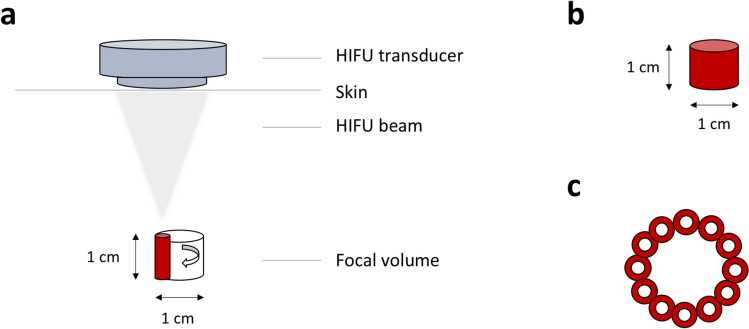
Objectives: Uterine fibroids often lead to symptoms that negatively impact health-related quality of life (HRQOL). High-intensity focused ultrasound (HIFU) has emerged as a promising noninvasive treatment for reducing fibroid size and symptoms. The Mirabilis system for ultrasound (US)-guided HIFU introduces a novel technique known as 'shell ablation'. This study evaluates the feasibility and efficacy of Mirabilis in a clinical setting, focusing on clinical outcomes.

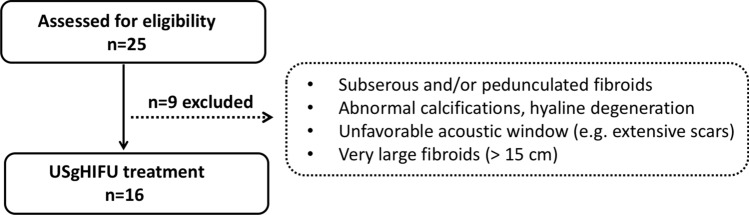
Materials and methods: Sixteen patients with 23 uterine fibroids were treated with the Mirabilis system. Follow-up assessments included US and MRI at baseline, 6 weeks, 3, 6 and 9 months, and 1 year after HIFU. Changes in symptoms and QOL were evaluated using the Uterine Fibroid Symptom and HRQOL Questionnaire.

Results: A significant reduction in fibroid volume was observed after HIFU (baseline 182.1 ± 49.3 ml; 1 year: 76.0 ± 37.9 ml, p < 0.001). The symptom severity score significantly declined (baseline 57.2 ± 3.8; 1 year: 30.2 ± 4.9, p < 0.001), correlating with a significant improvement in HRQOL (baseline 47.0 ± 3.9, 1 year: 71.8 ± 5.3, p < 0.001).

Conclusion: HIFU with the portable Mirabilis system is a feasible and safe noninvasive treatment for symptomatic uterine fibroids in an outpatient setting. This approach allows efficient and rapid ablation even for large fibroids, significantly reducing fibroid volume and symptoms.

Keywords: Fibroid-associated symptoms; High-intensity focused ultrasound; Mirabilis system; Mobile device; Shell ablation.


Conflict of interest statement

Declarations. Conflict of interests: The authors have no competing interests to declare that are relevant to the content of this article. Ethical approval: The study was conducted in accordance to the Declaration of Helsinki. It was approved by the local ethics committee of the University Hospital Bonn (No. 295/19), and it was registered in the German Clinical Trials Register (DRKS00020529). Written informed consent to participation was obtained from all patients.

Figures

Fig. 1
Fig. 1
Study consort diagram
Fig. 2
Fig. 2
Design of the Mirabilis system for rapid noninvasive HIFU ablation (Mirabilis Medical, Inc., Bothell, Washington, USA). The device consists of a portable cart base with a movable unit that is fixed to a mechanical arm and contains a diagnostic US device (BK Medical Model 2300; Analogic Corporation, Peabody, MA, USA) and a HIFU transducer. A touchscreen-based user interface displays diagnostic US images and allows controlling of the HIFU applicator and adjustment of treatment parameters. HIFU: high-intensity focused ultrasound, US: ultrasound
Fig. 3
Fig. 3
Volumetric shell ablation technique used by the Mirabilis system. a, b The extracorporal HIFU transducer generates a HIFU beam creating a focus with a length of 10 mm and a width of a few millimeters. Automatic rotation of the HIFU beam forms a cylinder-shaped unit volume. c Top view of multiple unit volumes that are created in the periphery of the target fibroid successively in shape of one or more rings to form a shell (maximum diameter: 5 cm, depth range: 3.5 – 9.5 cm). HIFU: high-intensity focused ultrasound
Fig. 4
Fig. 4
MRI images of a 40-year-old premenopausal patient with multiple fibroids. The patient presented with severe fibroid-associated symptoms (hypermenorrhea, pelvic pain and urinary urgency). There was no fibroid-specific previous therapy. The largest fibroid was located in the posterior wall of the uterus and was the main target of ablation. a Large target fibroid of the posterior wall of the uterus before HIFU treatment with a volume of 400.9 ml. The T2-weighted image shows that the target fibroid is partially isointense and partially hyperintense compared to myometrium indicating Funaki type 3. The contrast-enhanced T1-weighted image (c) shows an extensive enhancement of the fibroid (arrows). b The contrast-enhanced T1-weighted image shows an extensive enhancement of the fibroid. c One day after HIFU treatment. Contrast-enhanced T1-weighted MRI shows a large devascularized ablation volume with a NPV of 41.8%. HIFU: high-intensity focused ultrasound, MRI: magnetic resonance imaging, NPV: non-perfused volume
Fig. 5
Fig. 5
Average reduction of uterine fibroid volume (ml) after US-guided HIFU. Significance levels are indicated by the stars attached (** indicating p < 0.01, *** indicating p < 0.001). Standard errors were symmetrical (only positive error bars are shown). Significant volume regression was observed at each time point of follow-up between 6 weeks and 1 year compared to baseline. HIFU: high-intensity focused ultrasound, m: month, US: ultrasound, w: week, y: year
Fig. 6
Fig. 6
Changes in SSS and HRQOL mean scores of SSS and HRQOL after HIFU are presented. Significance levels are indicated by the stars attached (* indicating p < 0.05, ** indicating p < 0.01, *** indicating p < 0.001). Standard errors were symmetrical (positive error bars shown for HRQOL and negative bars shown for SSS). FU: Follow-up, HRQOL: health-related quality of life, m: month, SSS: symptom severity score, w: week, y: year

References

    1. Ea S, Cl C, Ra G et al (2017) Epidemiology of uterine fibroids a systematic review. BJOG An Int J Obstet Gynaecol. 124(10):1501–1512 - PubMed
    1. Baird Dd, Db D, Mc H et al (2003) High cumulative incidence of uterine leiomyoma in black and white women: ultrasound evidence. Am J Obstet Gynecol. 188(1):100–107 - PubMed
    1. Zimmermann A, Bernuit D, Gerlinger C et al (2012) Prevalence, symptoms and management of uterine fibroids: an international internet-based survey of 21,746 women. Bmc Women’s Health. 12:1–11 - PMC - PubMed
    1. As B, Reimer P, Matzko M et al (2014) The conservative and interventional treatment of fibroids. Dtsch Arztebl Int. 111(51–52):877–883. 10.3238/arztebl.2014.0877 - PMC - PubMed
    1. Donnez J, Dolmans M-M (2016) Uterine fibroid management: from the present to the future. Hum Reprod Update. 22(6):665–686. 10.1093/humupd/dmw023 - PMC - PubMed

Publication types

LinkOut - more resources