. 2025 Mar 10;21(3):e1012972.
doi: 10.1371/journal.ppat.1012972. eCollection 2025 Mar.

Unidirectional recruitment between MeCP2 and KSHV-encoded LANA revealed by CRISPR/Cas9 recruitment assay

Affiliations

Unidirectional recruitment between MeCP2 and KSHV-encoded LANA revealed by CRISPR/Cas9 recruitment assay

Ido Lavi et al. PLoS Pathog. .

Abstract

Kaposi's sarcoma-associated herpesvirus (KSHV, HHV-8) is associated with several human malignancies. During latency, the viral genomes reside in the nucleus of infected cells as large non-integrated plasmids, known as episomes. To ensure episome maintenance, the latency protein LANA tethers the viral episomes to the cell chromosomes during cell division. Directional recruitment of protein complexes is critical for the proper function of many nuclear processes. To test for recruitment directionality between LANA and cellular proteins, we directed LANA via catalytically inactive Cas9 (dCas9) to a repeat sequence to obtain easily detectable dots. Then, the recruitment of nuclear proteins to these dots can be evaluated. We termed this assay CRISPR-PITA for Protein Interaction and Telomere Recruitment Assay. Using this protein recruitment assay, we found that LANA recruits its known interactors ORC2 and SIN3A. Interestingly, LANA was unable to recruit MeCP2, but MeCP2 recruited LANA. Both LANA and histone deacetylase 1 (HDAC1) interact with the transcriptional-repression domain (TRD) and the methyl-CpG-binding domain (MBD) of MeCP2. Similar to LANA, HDAC1 was unable to recruit MeCP2. While heterochromatin protein 1 (HP1), which interacts with the N-terminal of MeCP2, can recruit MeCP2. We propose that available interacting domains force this recruitment directionality. We hypothesized that the tandem repeats in the SunTag may force MeCP2 dimerization and mimic the form of DNA-bound MeCP2. Indeed, providing only the tandem epitopes of SunTag allows LANA to recruit MeCP2 in infected cells. Therefore, CRISPR-PITA revealed the rules of unidirectional recruitment and allowed us to break this directionality.


Conflict of interest statement

The authors have declared that no competing interests exist.

Figures

Fig 1
Fig 1. Schematic illustration of CRISPR-PITA.
(A) Schematic illustration of dCas9-SunTag, sgRNA, and scFv-LANA or other protein of interest (scFv-pY). (B) SLK cells were transfected with dCas9-SunTag, scFv-LANA, and sgTelomere, as illustrated on the left of the images. An immunofluorescence assay detected LANA (red), and the nucleus was stained with DAPI (blue). Scale bar = 5 μm. Images are representatives of at least three independent experiments. (C) SLK cells were transfected with dCas9-SunTag, sgTelomere, and scFv-LANA or scFv-LANA oligomerization mutant, as illustrated on the left of the images. An immunofluorescence assay detected LANA (red) and TRF2 (white). The nucleus was stained with DAPI. Images are representatives of at least two independent experiments. (D) Schematic illustration of the CRISPR-PITA, targeting of scFv-LANA or any other protein of interest to the telomeres via dCas9. Then, the recruitment of other nuclear proteins to these dots is evaluated via immunostaining.
Fig 2
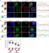
Fig 2. LANA recruits ORC2 and SIN3A but not MeCP2.
SLK cells were transfected with dCas9-SunTag, scFv-LANA, and sgTelomere expression plasmids, as indicated in the illustrations on the left. Immunofluorescence assays detected LANA (red) and ORC2 (A) mSin3a (B) or MeCP2 (C) (in green) cellular localization. The nucleus was stained with DAPI. Scale bar = 5 μm. Images are representatives of at least three independent experiments. The plots of the red, green, and blue pixel intensities along the white arrow (in the middle panels) are presented. (D) Pearson’s correlation coefficient was determined by ImageJ (JACoP Pluing) [62] for 15 cells in each treatment and presented as box and whiskers (min to max). Two-tailed t tests were performed (*, P ≤ 0.05; **, P ≤ 0.01; ***; P ≤ 0.001).
Fig 3
Fig 3. MeCP2 recruits LANA.
SLK cells were transfected with dCas9-SunTag, scFv-MeCP2, and sgTelomere in combination with GFP-LANA (A) or GFP (B), as illustrated on the left. An immunofluorescence assay detected scFv-MeCP2, or fluorescently labeled GFP-LANA and GFP. The nucleus was stained with DAPI. Scale bar = 5 μm. Images are representatives of at least three independent experiments. The plots of the red, green, and blue pixel intensities along the white arrow (in the middle panels) are presented. (C) SLK cells were transfected with FLAG-LANA N+C, FLAG-LANA C, or FLAG-LANA N, the same as in A. An immunofluorescence assay was performed to detect both scFv-MeCP2 and LANA. (D) Pearson’s correlation coefficient was determined for 15 cells in each treatment and presented as box and whiskers (min to max). Two-tailed t tests were performed (*, P ≤ 0.05; **, P ≤ 0.01; ***; P ≤ 0.001). (E) ChIP assay was performed on HEK293 cells to detect LANA (left panel) and MeCP2 (right panel) association with the ERBB2 promoter following recruitment with sgRNA to this locus.
Fig 4
Fig 4. MeCP2 T158M mutant fails to recruit LANA to heterochromatin in NIH3T3 cells but can recruit LANA when artificially tethered to DNA.
(A, B) NIH 3T3 cells were transfected with GFP-LANA and wt MeCP2, T158M mutant, or del-MBD. An immunofluorescence assay detected LANA (green) and MeCP2 (red). The heterochromatin foci in the nucleus of NIH3T3 mouse cells have a high intensity of DAPI (blue). Scale bar = 5 μm. Images are representatives of at least two independent experiments. The plots of the red, green, and blue pixel intensities along the white arrow (in the middle panels) are presented. (B) Pearson’s correlation coefficient was determined for 15 cells in each treatment and presented as box and whiskers (min to max). (C, D) rKSHV-infected iSLK cells (iSLK.Bac16) were transfected with dCas9-SunTag, scFv-MeCP2, and sgTelomere. An immunofluorescence assay was performed with rabbit anti-HA and rat anti-LANA and anti-rabbit Alexa Flour 555 and anti-rat Alexa Flour 647 antibodies, to detect scFv-MeCP2 and LANA dots in infected cells. In this case, the signal from anti-Rat Alexa Flour 647 antibody was pseudo-color to green to keep it consistent with the other CRISPR-PITA experiments. (D) Pearson’s correlation coefficient was determined same as in B for the experiment in C. Two-tailed t-tests were performed (*, P ≤ 0.05; **, P ≤ 0.01; ***; P ≤ 0.001, ****; P ≤ 0.0001).
Fig 5
Fig 5. MeCP2 recruits HDAC1, while HDAC1 cannot recruit MeCP2.
(A) As illustrated on the left, SLK cells were transfected with dCas9-SunTag, scFv-MeCP2, and sgTelomere. An immunofluorescence assay detected HDAC1 (green) and scFv-MeCP2 (red). (B) SLK cells were transfected the same as in A but with scFv-HDAC1. An immunofluorescence assay detected MeCP2 (green) and scFv-HDAC1 (red). The nucleus was stained with DAPI. Scale bar = 5 μm. Images are representatives of at least two independent experiments. The plots of the red, green, and blue pixel intensities along the white arrow (in the middle panels) are presented. (C) Pearson’s correlation coefficient was determined for 15 cells in each treatment and presented as box and whiskers (min to max). Two-tailed t-tests were performed (****; P ≤ 0.0001).
Fig 6
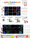
Fig 6. The availability of interacting domains determines recruitment directionality.
(A) schematic presentation of MeCP2 and the interaction domains with LANA, HDAC1, and HP1α. Two Rett syndrome mutants are indicated above. (B) SLK cells were transfected with dCas9-SunTag, scFv-HP1α, and sgTelomere, as illustrated on the left. An immunofluorescence assay detected MeCP2 (green) and scFv-HP1α (red). The nucleus was stained with DAPI. Scale bar = 5 μm. Images are representatives of at least two independent experiments. (C) Pearson’s correlation coefficient was determined for 15 cells in each treatment and presented as box and whiskers (min to max). Two-tailed t-tests were performed. Schematic illustrations of the model for free (D) DNA-bound MeCP2 (E) and SunTag scFv-MeCP2 (F) are presented. (G) iSLK-infected cells (iSLK.Bac16) were transfected with free SunTag and scFv-MeCP2. Then, an immunofluorescence assay was performed to detect scFv-MeCP2 (red) and LANA (yellow). GFP is a marker of infected cells. The plots of the red, yellow, green, and blue pixel intensities along the white arrow (in the middle panels) are presented. (H) Pearson’s correlation coefficient was determined for 15 cells in each treatment and presented as box and whiskers (min to max). (I) Schematic presentation of the experiment in G and its conclusions.

References

    1. Henke-Gendo C, Schulz TF. Transmission and disease association of Kaposi’s sarcoma-associated herpesvirus: recent developments. Curr Opin Infect Dis. 2004;17(1):53–7. doi: 10.1097/00001432-200402000-00011 - DOI - PubMed
    1. Chang Y, Cesarman E, Pessin MS, Lee F, Culpepper J, Knowles DM, et al.. Identification of herpesvirus-like DNA sequences in AIDS-associated Kaposi’s sarcoma. Science. 1994;266(5192):1865–9. doi: 10.1126/science.7997879 - DOI - PubMed
    1. Kedes DH, Lagunoff M, Renne R, Ganem D. Identification of the gene encoding the major latency-associated nuclear antigen of the Kaposi’s sarcoma-associated herpesvirus. J Clin Invest. 1997;100(10):2606–10. doi: 10.1172/JCI119804 - DOI - PMC - PubMed
    1. Rainbow L, Platt GM, Simpson GR, Sarid R, Gao SJ, Stoiber H, et al.. The 222- to 234-kilodalton latent nuclear protein (LNA) of Kaposi’s sarcoma-associated herpesvirus (human herpesvirus 8) is encoded by orf73 and is a component of the latency-associated nuclear antigen. J Virol. 1997;71(8):5915–21. doi: 10.1128/JVI.71.8.5915-5921.1997 - DOI - PMC - PubMed
    1. Ye F, Lei X, Gao S-J. Mechanisms of Kaposi’s Sarcoma-Associated Herpesvirus Latency and Reactivation. Adv Virol. 2011;2011:193860. doi: 10.1155/2011/193860 - DOI - PMC - PubMed

MeSH terms