. 2025 Mar 26;147(12):10188-10202.
doi: 10.1021/jacs.4c15223. Epub 2025 Mar 10.

Highly Specific Miniaturized Fluorescent Monoacylglycerol Lipase Probes Enable Translational Research

Affiliations

Highly Specific Miniaturized Fluorescent Monoacylglycerol Lipase Probes Enable Translational Research

Axel Hentsch et al. J Am Chem Soc. .

Abstract

Monoacylglycerol lipase (MAGL) is the pivotal catabolic enzyme responsible for signal termination in the endocannabinoid system. Inhibition of MAGL offers unique advantages over the direct activation of cannabinoid receptors in treating cancer, metabolic disorders, and inflammatory diseases. Although specific fluorescent molecular imaging probes are commonly used for the real-time analysis of the localization and distribution of drug targets in cells, they are almost invariably composed of a linker connecting the pharmacophore with a large fluorophore. In this study, we have developed miniaturized fluorescent probes targeting MAGL by incorporating a highly fluorescent boron-dipyrromethene (BODIPY) moiety into the inhibitor structure that interacts with the MAGL active site. These miniaturized fluorescent probes exhibit favorable drug-like properties such as high solubility and permeability, picomolar potency for MAGL across various species, and high cell selectivity and specificity. A range of translational investigations were conducted, including cell-free fluorescence polarization assays, fluorescence-activated cell sorting analysis, and confocal fluorescence microscopy of live cancer cells, live primary neurons, and human-induced pluripotent stem cell-derived brain organoids. Furthermore, the application of red-shifted analogs or 18F positron emission labeling illustrated the significant versatility and adaptability of the fluorescent ligands in various experimental contexts.


Conflict of interest statement

The authors declare no competing financial interest.

Figures

Scheme 1
Scheme 1. Miniaturization Approach by the Partial Replacement of the Monoacylglycerol Lipase (MAGL) Pharmacophore with the Boron-Dipyrromethene (BODIPY) Fluorophore
Comparison of the miniaturized fluorescent probe 1 with the highly lipophilic, classically constructed fluorescent probe LEI-463. The miniaturized probes exhibit highly drug-like properties. Their versatility is showcased in various experimental translational settings of clinical relevance, such as activity-based profiling, live cell imaging, flow cytometry and radiolabeling.
Scheme 2
Scheme 2. Synthetic Access to BODIPY MAGL Probes
(A) General synthetic approach for 8-methylene substituted fluorescent BODIPY MAGL probes involves two key connection steps. The first step is the formation of an amide or urea (non-covalent inhibitor) or carbamate (covalent inhibitor) to connect the headgroup to the cyclic amine and the second step is a Liebeskind–Srogl cross-coupling (LSCC) reaction to attach the BODIPY moiety. Reaction conditions: (a) LiTMP, CH2(Bpin)2, THF, –78 °C to rt, 18 h. (b) TFA/DCM 1:4, rt, 3 h. (c) Various coupling reactions using activated carbamates (see SI for further information). (d) H2O, NH4OAc, NaIO4, acetone, rt, 16–24 h. (e) Cu(I)TC, Pd2(dba)3, TFP, THF, 55 °C, 10–90 min. (f) Et3SiH, Pd/C, MeOH, 0 °C to rt, 10–120 min. (g) PhNH2, t-BuONO, MeCN, 40 °C, 16 h. (h) Neat pyrrole, MW, 150 °C, 2–3 h. (B) Synthesis of irreversible, covalent compound 1. Reaction conditions and yields are indicated in the synthetic sequence. (C) Overview of the examined SAR (for complete structures and SAR data see SI, Figure S1).
Scheme 3
Scheme 3. Overview of the Most Active Miniaturized Fluorescent Probes
Figure 1
Figure 1
Co-crystal structure of miniaturized fluorescent probe 5 in complex with hMAGL (PDB: 8RVF, resolution = 1.43 Å). (A) Binding mode of compound 5 in hMAGL with water-mediated and direct protein–ligand hydrogen bond interactions indicated by dashed lines (blue for direct binding, red for water-mediated interactions). Key residues for hydrogen bonding, including the oxyanion hole (Ala51, Met123) and catalytic Ser122, are highlighted in stick representation. Additional water molecules have been removed for clarity of view. (B) Close-up view of the boron-dipyrromethene (BODIPY) environment buried in the hMAGL-binding site with indicated key interactions. Short nonbonded contacts (d ≤ 4.0 Å), either in direct contact with MAGL residues (blue) or mediated by water (red), are shown by dashed lines.
Figure 2
Figure 2
(A) Multiplex activity-based protein profiling (ABPP) assay was used to label various serine hydrolases in the membrane and cytosolic fraction of the mouse brain proteome (MBP). Two MAGL bands at approximately 35 kDa were selectively blocked by 1 μM of the respective reversible probes 4, 4a, 5, 5a. Both panels depict the same representative SDS-PAGE at the respective fluorescent channel settings. (B) Dose-dependent labeling of the two MAGL splicing variants in MBP with the four irreversible probes 2, 1, 2a, 1a. (C) Irreversible probes as MAGL-selective ABPP probes for target-occupancy assessment in patient-derived human peripheral blood monocytes (PBMCs) at 100 nM. The MAGL fluorescence signal was dose-dependent to MAGL inhibitor PF. Coomassie protein staining as a loading control.
Figure 3
Figure 3
Cell-free fluorescence polarization (FP) and cellular nanoBRET competition assays using the reversible noncovalent fluorescent probes 5 and 4a as tracers. (A) Fluorescence polarization dose–response titration of reversible probe 5 (20 nM) with purified hMAGL. (B) Three MAGL-specific inhibitors were assessed in a competitive FP assay with 5 (20 nM) and hMAGL (50 nM). (C) Competition nanoBRET assay in live HEK293 cells expressing MAGL-Nluc fusion protein and reversible probe 4a (100 nM). All experiments were performed at least in triplicate. Mean values are shown.
Figure 4
Figure 4
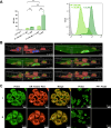
(A) Mean fluorescence intensity (MFI) in the flow cytometry analysis of HT-29 cells with probe 1. Data was confirmed in two independent experiments, triplicate determination per condition. A one-way ANOVA test was performed. ** indicates p < 0.005. Histogram of HT-29 cells: negative control (gray), incubation with 1 (5 μM) for 60 min with preincubation of MAGL inhibitor, PF (100 μM; bright green), without blocking (dark green). (B) Uptake process of probe 1 in live HT-29 cells without washing. Three-dimensional confocal microscopic image composite with staining of nuclei (Hoechst; blue), cell membrane (CellMask-A647; Invitrogen; red), and probe 1 (green). Images were acquired using a 63× water immersion objective. Each of the z-stacks consists of 50 confocal planes at a plane distance of 0.5 μm. One field of view is illustrated (200 μm × 200 μm × 25 μm). (C) Confocal live cell microscopy of HT-29 cells revealed the colocalization of MAGL probes 1 and 5 (150 nM, FITC channel) and endoplasmic reticulum ER-tracker (ER-Tracker Red; Invitrogen, Cy3 channel). Preincubation with PF (10 μM) blocks the fluorescence signal.
Figure 5
Figure 5
Confocal imaging of primary neuronal cultures and human induced pluripotent stem cell (hiPSC)-derived brain organoids using covalent probe 1. (A) Left: confocal images of the live dissociated primary hippocampal neuron culture at day-in vitro 14–21. Cells were treated with 1 (1 μM) at 37 °C for 15 min; negative controls were treated with 10 μM MAGL inhibitor PF for 2 h prior to MAGL labeling. Neuronal processes (dashed line) and potential synapses (insets) are highlighted; right: quantification of mean fluorescence intensity (MFI) of the signal of 1 in neurons with or without 10 μM PF pretreatment (open circles represent n = 4 images per condition, two-tailed Student t test). (B) Representative confocal images of human induced pluripotent stem cell (hiPSC)-derived brain organoids incubated with 5 μM probe 1 for 2 h after 83 d of organoid maturation, subsequent fixation, cryosectioning, and staining of nuclei (Hoechst) and neurons (MAP2). Subpanel row (a): overview with an indication of regions for subpanels rows (b, c). Arrows indicate several MAP2-positive neurons (subpanel b) and non-neuronal cells, MAP2-negative; subpanel (c) with signal for probe 1.
Figure 6
Figure 6
(A) Synthetic conversion of probe 2 into a bimodal fluorescent positron emission tomography (PET) probe [18F]-2. (B) In vitro autoradiography of wild-type and MAGL knockout mouse brain sections with [18F]-2 (45 nM, 8 GBq/μmol).

References

    1. Dinh T. P.; Carpenter D.; Leslie F. M.; Freund T. F.; Katona I.; Sensi S. L.; Kathuria S.; Piomelli D. Brain Monoglyceride Lipase Participating in Endocannabinoid Inactivation. Proc. Natl. Acad. Sci. U.S.A. 2002, 99 (16), 10819–10824. 10.1073/pnas.152334899. - DOI - PMC - PubMed
    1. Stella N.; Schweitzer P.; Piomelli D. A Second Endogenous Cannabinoid That Modulates Long-Term Potentiation. Nature 1997, 388, 773–778. 10.1038/42015. - DOI - PubMed
    1. Kano M.; Ohno-Shosaku T.; Hashimotodani Y.; Uchigashima M.; Watanabe M. Endocannabinoid-Mediated Control of Synaptic Transmission. Physiol. Rev. 2009, 89 (1), 309–380. 10.1152/physrev.00019.2008. - DOI - PubMed
    1. Nomura D. K.; Long J. Z.; Niessen S.; Hoover H. S.; Ng S. W.; Cravatt B. F. Monoacylglycerol Lipase Regulates a Fatty Acid Network That Promotes Cancer Pathogenesis. Cell 2010, 140 (1), 49–61. 10.1016/j.cell.2009.11.027. - DOI - PMC - PubMed
    1. Nomura D. K.; Morrison B. E.; Blankman J. L.; Long J. Z.; Kinsey S. G.; Marcondes M. C. G.; Ward A. M.; Hahn Y. K.; Lichtman A. H.; Conti B.; Cravatt B. F. Endocannabinoid Hydrolysis Generates Brain Prostaglandins That Promote Neuroinflammation. Science 2011, 334 (6057), 809–813. 10.1126/science.1209200. - DOI - PMC - PubMed

MeSH terms

LinkOut - more resources