. 2025 Mar 10;16(1):2368.
doi: 10.1038/s41467-025-57488-0.

Berberine-inspired ionizable lipid for self-structure stabilization and brain targeting delivery of nucleic acid therapeutics

Affiliations

Berberine-inspired ionizable lipid for self-structure stabilization and brain targeting delivery of nucleic acid therapeutics

Xufei Bian et al. Nat Commun. .

Abstract

Lipid nanoparticles have shown success in targeting major organs such as the liver, spleen, and lungs, but crossing the blood-brain barrier (BBB) remains a major challenge. Effective brain-targeted delivery systems are essential for advancing gene therapy for neurological diseases but remain limited by low transport efficiency and poor nucleic acid stability. Here, we report a library of ionizable lipids based on the tetrahydroisoquinoline structure of protoberberine alkaloids, designed to improve BBB penetration via dopamine D3 receptor-mediated endocytosis. These nanoparticles offer three key advantages: enhanced brain uptake, improved nucleic acid stability through poly(A) self-assembly, and minimal immunogenicity with inherent neuroprotective properties. In murine models, they demonstrate therapeutic potential in Alzheimer's disease, glioma, and cryptococcal meningitis. This berberine-inspired delivery system integrates precise receptor targeting with nucleic acid stabilization, offering a promising platform for brain-targeted therapeutics.


Conflict of interest statement

Competing interests: The authors declare no competing interests.

Figures

Fig. 1
Fig. 1. Design and mechanism of berberine-inspired lipidoid nanoparticles (BE) for targeted brain delivery.
a Construction of a three-dimensional combinatorial synthesis library and formation of BE@RNA complexes through berberine derivative-poly(A) affinity and self-assembly properties. b Blood-brain barrier penetration mechanism through dopamine D3 receptor (D3R)-mediated transport, enabling targeted therapeutic delivery for neurodegenerative disorders, including Alzheimer’s disease.
Fig. 2
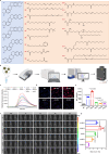
Fig. 2. Design and characterization of tetrahydroisoquinoline alkaloid-based ionizable lipids.
a Chemical structures of lipid building blocks comprising five amine heads and 15 alkylated tails. b Optimization workflow for BE lipidoid molecule development, involving 27 formulations derived from 75 novel ionizable compounds. (Created with BioRender.com) c Affinities of various ionizable lipidoid molecules for poly(A) measured using fluorescence spectroscopy. d, e Cellular uptake of LNPs assessed by fluorescence co-localization studies and flow cytometry analysis. LNPs were labeled DiD (red). Nuclei were stained with Hoechst 33342 (blue). Scale bar, 20 µm. f In vivo imaging of mice after injection of LNP@DiR at 0.5, 1, 2, 4, and 8 h. g Ex vivo fluorescence distribution showing brain-to-liver ratio at 1 h. Data are presented as mean ± SD (n = 3 independent experiments). Statistical significance was determined by two-tailed unpaired Student’s t test (two groups) or one-way ANOVA with Dunnett’s multiple comparison tests (multiple groups); **P < 0.01, ***P < 0.001, ****P < 0.0001. Source data are provided as a Source Data file.
Fig. 3
Fig. 3. In vitro characterization of BE-based LNPs containing ionizable lipid A2-B13.
a Gel retardation assay of BE@siRNA at LNP/siRNA weight ratios from 0.2 to 20 to evaluate complex stability. b Size distribution analysis by dynamic light scattering. c Transmission electron microscopy (TEM) image showing particle morphology of BE@siRNA. Scale bar, 100 nm. d Cellular uptake comparison between BE@FAM-siRNA and MC3@FAM-siRNA by fluorescence microscopy. Scale bar, 25 µm. e High-magnification imaging of subcellular localization of BE@siRNA and MC3@siRNA. FAM channel shows BE@FAM-siRNAs (green). LysoTracker channel shows lysosome (red). Nuclei were counter stained with Hoechst 33342 (blue). Scale bar, 5 µm. f Analysis of BE-mediated endocytosis mechanisms. g, h LNP@DiD penetration across an in vitro blood-brain barrier (BBB) model using transwell assays. LNPs were labeled DiD (red). Nuclei were counter stained with Hoechst 33342 (blue). Scale bar, 1000 µm. Data are presented as mean ± SD (n = 3 independent experiments). Statistical significance was determined by two-tailed unpaired Student’s t test (two groups) or one-way ANOVA with Dunnett’s multiple comparison tests (multiple groups); ***P < 0.001; NS not significant. Data are representative of three (c, d) and two (e) independent experiments with similar results. Source data are provided as a Source Data file.
Fig. 4
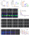
Fig. 4. Brain-targeting evaluation of BE-ST nanoparticles.
a Cellular uptake analysis by flow cytometry following DRD3 knockdown with siRNA. b Zeta potential measurements of BE complexed with different organic acids. c Brain-to-liver fluorescence distribution ratio of BE formulated with various organic acids by ex vivo imaging. d In vivo and ex vivo imaging of LNP@DiR distribution 1 h post-injection of BE-organic acid complexes. e Plasma concentration-time profiles of BE and BE-ST (1:5 w/w) in SD rats following intravenous administration. f Time-course in vivo imaging of LNP@DiR distribution at 0.5, 1, 2, 4, and 8 h post-injection. g Ex vivo brain imaging at 1, 2, 4, and 8 h post-administration. h Quantitative analysis of brain fluorescence intensity over time. Data are presented as mean ± SD (n = 3 independent experiments). Statistical significance was determined by two-tailed unpaired Student’s t test; *P < 0.05, **P < 0.01, ***P < 0.001, ****P < 0.0001; NS not significant. Source data are provided as a Source Data file.
Fig. 5
Fig. 5. BE(-ST)@siBACE1 therapy improves cognitive function in APP/PS1 mice.
a Experimental timeline showing treatment regimen of APP/PS1 and wild-type (WT) mice receiving tail vein injections of LNP@siBACE1 or PBS every 2 days for seven cycles, followed by Morris water maze (MWM) testing and tissue collection. be MWM probe test results: b Representative swimming trajectories during platform search. c Time spent in target quadrant as percentage of total time. d Platform location crossing frequency. e Mean swimming speed across groups. Data are presented as mean ± SD (n = 6 biologically independent animals). Statistical significance was determined by two-tailed unpaired Student’s t test; *P < 0.05, **P < 0.01, ***P < 0.001. Source data are provided as a Source Data file.
Fig. 6
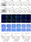
Fig. 6. Therapeutic synergy of BE(-ST) and siBACE1 in modulating Alzheimer’s disease (AD) hallmarks in APP/PS1 mice.
a Representative western blot images showing BACE1, phosphorylated tau (p-tau), and GSK3β levels in the hippocampus and cortex of BE(-ST)@siBACE1-treated APP/PS1 mice, control APP/PS1 groups, and wild-type (WT) mice. b Quantification of western blot for BACE1, p-tau, and phosphorylated GSK3β (p-GSK3β), normalized to GAPDH. The samples derive from the same experiment and that blots were processed in parallel. c Representative confocal laser scanning microscopy images showing amyloid-β (Aβ) plaque burden in the hippocampus and cortex of APP/PS1 and WT mice. Aβ plaques (green) and nuclei (Hoechst 33342, blue). Scale bars: top row, 500 µm; bottom row, 250 µm. d Nissl staining of brain sections 14 days post-treatment. Scale bar, 50 µm. ELISA results for complement activation-related pseudoallergy (CARPA) markers: e C5b9, f C3a, and g monocyte chemoattractant protein (MCP-1) in serum. Data are means ± SD (n = 3 biologically independent samples). Statistical significance was determined by two-tailed unpaired Student’s t test; *P < 0.05, **P < 0.01, ***P < 0.001, ****P < 0.0001. Data in c, d are representative of two independent experiments with similar results. Source data are provided as a Source Data file.
Fig. 7
Fig. 7. BE-ST enables efficient delivery of small-molecule and macromolecular drugs in vivo.
a Experimental timeline for cryptococcal meningitis treatment in mice using amphotericin B (AMB). b, c In vivo fluorescence imaging and quantification. Data are presented as mean ± SD (n = 5 biologically independent animals). d Brain tissue fungal colony burden quantification. Data are presented as mean ± SD (n = 6 biologically independent animals). e Kaplan-Meier survival analysis (n = 10 biologically independent animals). f Fluorescence microscopy of eGFP mRNA in vitro transfection. Scale bar, 25 µm. g Three-dimensional visualization of polyA-A2-B13 binding site interactions (PDB ID: 3GIB). h Agarose gel electrophoresis showing BE-loaded mRNA stability. i Three-dimensional hyalinization microscopy of brain tissue following 10 µg eGFP mRNA administration. Scale bar, 1500 µm. Statistical significance was determined by two-tailed unpaired Student’s t test; *P < 0.05, **P < 0.01, ***P < 0.001, ****P < 0.0001. Data are representative of three (f, h) and two (i) independent experiments with similar results. Source data are provided as a Source Data file.

References

    1. Chung, J. E. et al. Self-assembled micellar nanocomplexes comprising green tea catechin derivatives and protein drugs for cancer therapy. Nat. Nanotechnol.9, 907–912 (2014). - PMC - PubMed
    1. Shin, M. et al. Targeting protein and peptide therapeutics to the heart via tannic acid modification. Nat. Biomed. Eng.2, 304–317 (2018). - PubMed
    1. Wang, X. et al. Identification and construction of a novel biomimetic delivery system of paclitaxel and its targeting therapy for cancer. Signal Transduct. Target Ther.6, 33 (2021). - PMC - PubMed
    1. Fu, S. & Yang, X. Recent advances in natural small molecules as drug delivery systems. J. Mater. Chem. B11, 4584–4599 (2023). - PubMed
    1. Yan, Y. et al. Research progress on antibacterial activities and mechanisms of natural alkaloids: a review. Antibiotics10, 318 (2021). - PMC - PubMed

LinkOut - more resources