. 2025 Mar 10;16(1):2394.
doi: 10.1038/s41467-025-57628-6.

In vivo prime editing rescues photoreceptor degeneration in nonsense mutant retinitis pigmentosa

Affiliations

In vivo prime editing rescues photoreceptor degeneration in nonsense mutant retinitis pigmentosa

Yidian Fu et al. Nat Commun. .

Abstract

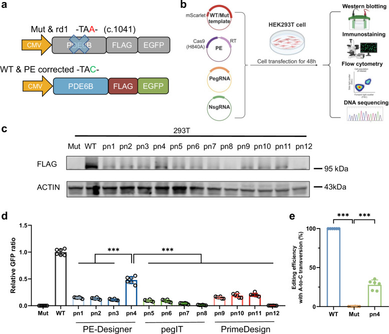
The next-generation gene editing tool, prime editing (PE), is adept at correcting point mutations precisely with high editing efficiency and rare off-target events and shows promising therapeutic value in treating hereditary diseases. Retinitis pigmentosa (RP) is the most common type of inherited retinal dystrophy and is characterized by progressive degeneration of retinal photoreceptors and, consequently, visual decline. To date, effective treatments for RP are lacking. Herein, a PE system is designed to target the PDE6B Y347X mutation in the rd1 mouse strain, a preclinical RP model. We screen and develop the PE system with epegRNA and RTΔRnH, which is delivered via dual-AAV in vivo with an editing efficiency of 26.47 ± 13.35%, with negligible off-target effects confirmed by AID-Seq and PE-tag. Treatment with the PE system in vivo greatly restores PDE6B protein expression and protects rod cells from degeneration. Mouse behavioural experiments also show that compared with no treatment, prime editing inhibits vision deterioration in littermate rd1 mice. This study provides a therapeutic opportunity for the use of PE to correct mutated RPs at the genomic level.


Conflict of interest statement

Competing interests: D.R.L. is a co-founder and consultant for Prime Medicine, Beam Therapeutics, Pairwise Plants, and nChroma Bio, and owns equity in these companies. The other authors declare no competing interests.

Figures

Fig. 1
Fig. 1. Genetic background of RP and PE system design for the Pde6b mutation locus.
a Genetic background of RP. b Schematic representation of the Pde6b nonsense mutation in rd1 mice. The C-to-A mutation in exon 7 of the Pde6b locus induces a stop codon and inhibits normal expression of the PDE6B protein. Prime editing was designed to correct the A base to the C base. c Flowchart of pegRNA and nsgRNA design. The sequence of exon 7 of the mouse Pde6b gene was input into three websites (PE-Designer, pegIT and PrimeDesign). Among the output results, four pegRNA and nsgRNA pairs were randomly selected from each website. The corresponding pairs were named pn1-12. The sequence of each pair was illustrated below.
Fig. 2
Fig. 2. In vitro screening of pegRNAs and nsgRNAs targeting Pde6b mutation.
a Schematic representation of the reporter system. The template carrying exon 7 of the Pde6b gene with (mut template) or without the PDE6B Y347X mutation (WT template) was followed by sequences encoding a 3xFlag and EGFP. After removal of the stop codon via prime editing, the Flag protein tag and EGFP signal were detected. b Flowchart of the in vitro screening for efficient guide RNAs. 293 T cells were co-transfected with plasmids expressing the WT/mut template (containing mScarlet for indicating transfection efficiency) and PE system (prime editors, pegRNA and nsgRNA). The pn1-12 plasmids were individually added to independent groups. After transfection for 48 h, the cells were harvested for Western blotting, immunostaining, flow cytometry and DNA amplicon sequencing. Created in BioRender. c Western blot analysis of PDE6B-FLAG expression in 293 T cell lysates after 48 h of transfection. Uncropped blots in Source Data. d Relative GFP ratio was quantified from Immunofluorescence images of in vitro screening. Representative data are presented as the mean ± SD, n = 6 biologically independent replicates. One-way ANOVA with Tukey’s multiple comparisons test was used. ***p < 0.001. e Editing efficiency of the prime editing system for pn4. Representative data are presented as the mean ± SD, n = 6 biologically independent replicates. One-way ANOVA with Tukey’s multiple comparisons test was used. ***p < 0.001.
Fig. 3
Fig. 3. PE system precisely corrected the PDE6B Y347X mutation in rd1 mice in vivo.
a Schematic diagram of the PE system delivered by dual-AAV. Cas9(H840A) was split at site 713, at which the N-terminus followed by the epegRNA and nsgRNA was packaged in one AAV, whereas the C-terminus linked to the RTΔRnH domain was packaged in the other AAV. A trans-splicing intein system was employed to reconstitute the two AAVs into the full-length PE. b Timeline of the in vivo AAV-PE treatment. One-week-old rd1 mice were subjected to subretinal injection of the dual-AAV system. Three weeks after injection, PE-treated rd1 mice were subjected to a series of functional assays for vision evaluation. Then, the PE-treated rd1 mice were sacrificed at five weeks of age, and their eyeballs were collected for molecular biology and morphological analyses. Untreated littermates and WT mice of the same age were used as negative and positive controls, respectively. Created in BioRender. c The editing efficiency of the PE system in vivo. Representative data are presented as the mean ± SD, n = 6. One-way ANOVA with Tukey’s multiple comparisons test. *p < 0.05; ***p < 0.001. d Western blot analysis of PDE6B expression in mouse eyeball lysates after treatment at P35. Uncropped blots in Source Data. e Representative immunofluorescence image of the rod photoreceptor layer marked with rhodopsin (red) at P35. ONL, outer nuclear layer; INL, inner nuclear layer; OS, outer segment. Scale bar = 25 μm. f Quantification of OS thickness in WT, rd1, and PE-edited mice at P35. The means ± SDs are shown, n = 6. One-way ANOVA with Tukey’s multiple comparisons test was used for comparisons. **P < 0.01; ***p < 0.001. g Representative retinal section of WT, rd1, and PE-edited mice with H&E staining at P35. ONL, outer nuclear layer; INL, inner nuclear layer; and GCL, ganglion cell layer. Above scale bar = 100 μm. Below scale bar = 50 μm. h Quantification of ONL thickness in WT, rd1, and PE-edited mice at P35. One-way ANOVA with Tukey’s multiple comparisons test was used for comparisons. The means ± SDs are shown, n = 6. ***p < 0.001.
Fig. 4
Fig. 4. Assessment of the off-target effects of the PE system in rd1 mice.
a Schematic representation of AID-seq for assessing potential off-target sites. Created in BioRender. b Potential off-target sites detected by AID-seq. c Schematic representation of potential off-target sites for peg4. d Schematic representation of potential off-target sites for nsg4. e Amplicon sequencing assessment of potential off-target sites induced by the PE system. Representative data are presented as the mean ± SD. Generally, n = 6 replicates (otherwise n = 5). Two-tailed unpaired t tests were used. ns, no significance.
Fig. 5
Fig. 5. RNA-seq analysis of genes relevant to phototransduction after PE correction.
a KEGG enrichment analysis: downregulated phototransduction pathway (rd1 versus the WT group). Benjamini Hochberg were used. FDR < 0.05. b KEGG enrichment analysis: upregulated phototransduction pathway (the PE-edited group versus the rd1 group). Benjamini Hochberg were used. FDR < 0.05. c Volcano plot of genes differentially expressed between rd1 and WT mice. The red dots are upregulated genes, whereas the blue dots are downregulated genes. (t tests were used; |log2(fold change)|> 1 and p < 0.05). Genes relevant to the phototransduction pathway are highlighted. d Volcano plot of differentially expressed genes between PE-edited and rd1 mice. (t tests were used; |log2(fold change)|> 1 and p < 0.05). e The mRNA expression levels of genes relevant to the phototransduction pathway were detected in RNA-seq samples by RT‒qPCR. Representative data are presented as the mean ± SEM. Generally, n = 6 replicates (otherwise n = 4). Two-tailed unpaired t tests were used. ns, not significant; *p < 0.05; **p < 0.01.
Fig. 6
Fig. 6. AAV-PE restored visual function and improved vision-guided behavior in rd1 mice.
a Representative images of eye constriction in WT, rd1 and PE-treated mice. b Quantification of pupil constriction proportions in WT, rd1 and PE-treated mice. Representative data are presented as the mean ± SD, n = 6. One-way ANOVA with Tukey’s multiple comparisons test was used for comparisons. WT group vs rd1 group, p < 0.001; rd1 group vs PE-treated group, p < 0.001; PE-treated group vs WT group, p = 0.002. **p < 0.01; ***p < 0.001. c Schematic representation of the light–dark transition test. Mice could shuttle between the bright and dark boxes through the aperture. Created in BioRender. d Quantification of the amount of time spent in the dark zone in the WT, rd1 and PE-treated groups. Representative data are presented as the mean ± SD, n = 6. One-way ANOVA with Tukey’s multiple comparisons test was used for comparisons. WT group vs rd1 group, p < 0.001; rd1 group vs PE-treated group, p = 0.004; PE-treated group vs WT group, p = 0.046. *p < 0.05; **p < 0.01; ***p < 0.001. e Schematic representation of the visual cliff test. Created in BioRender. f Representative traveling trajectories (above) and heatmap recordings for time spent in distinct regions of the whole platform (below) in the WT, rd1 and PE-treated groups. g Quantification of the amount of time spent on the cliff side by WT, rd1 and PE-treated mice. Representative data are presented as the mean ± SD, n = 6. One-way ANOVA with Tukey’s multiple comparisons test was used for comparisons. WT group vs rd1 group, p < 0.001; rd1 group vs PE-treated group, p = 0.036; PE-treated group vs WT group, p = 0.015. *p < 0.05; ***p < 0.001.
Fig. 7
Fig. 7. Flowchart of prime editing application in treating retinitis pigmentosa.
Schematic representation of the flowchart for prime editing application in treating retinitis pigmentosa. The pegRNA and nsgRNA were first designed and cloned for the target site. Then in vitro transfection was performed to screen out the most efficient pairs of pegRNA and nsgRNA. Deep sequencing confirmed the editing efficiency in vitro. Next, AAV delivering PE system was applied in vivo through subretinal injection. In vivo editing was confirmed by deep sequencing and rescued retinal degeneration. Created in BioRender.

References

    1. Anzalone, A. V. et al. Search-and-replace genome editing without double-strand breaks or donor DNA. Nature576, 149–157 (2019). - PMC - PubMed
    1. Fu, Y. et al. Prime editing: current advances and therapeutic opportunities in human diseases. Sci. Bull.68, 3278–3291 (2023). - PubMed
    1. Jang, H. et al. Application of prime editing to the correction of mutations and phenotypes in adult mice with liver and eye diseases. Nat Biomed Eng, 10.1038/s41551-021-00788-9 (2021). - PubMed
    1. Jiang, T., Zhang, X. O., Weng, Z. & Xue, W. Deletion and replacement of long genomic sequences using prime editing. Nat. Biotechnol.40, 227–234 (2022). - PMC - PubMed
    1. Happi Mbakam, C. et al. Prime editing optimized RTT permits the correction of the c.8713C>T mutation in DMD gene. Mol. Ther. Nucleic Acids30, 272–285 (2022). - PMC - PubMed

MeSH terms

Substances

LinkOut - more resources