. 2025 Mar 10;15(1):8257.
doi: 10.1038/s41598-025-90689-7.

Diversity of ER-positive and HER2-negative breast cancer stem cells attained using selective culture techniques

Affiliations

Diversity of ER-positive and HER2-negative breast cancer stem cells attained using selective culture techniques

Satoshi Sueoka et al. Sci Rep. .

Abstract

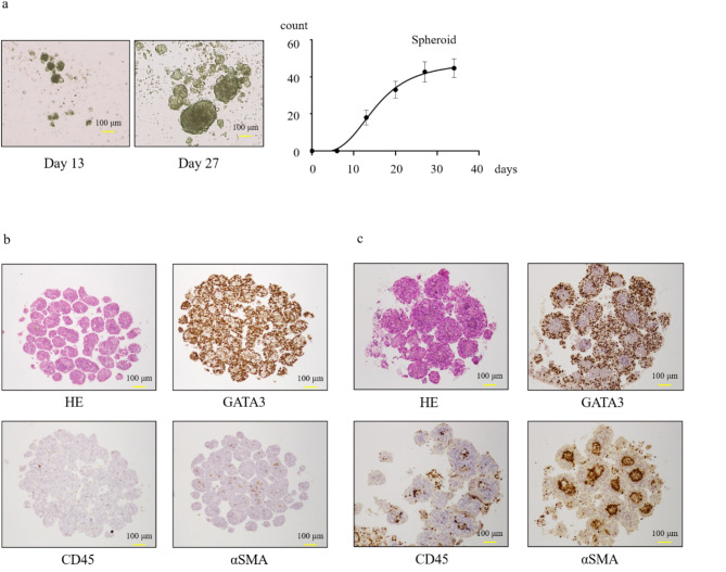
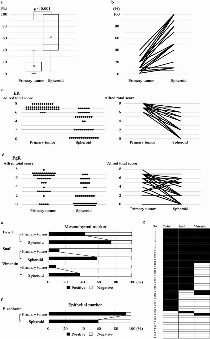
Breast cancer stem cells are a promising therapeutic target in cancer. We explored breast cancer stem cell diversity and establish a methodology for selectively culturing breast cancer stem cells. We collected breast cancer tissues from surgical samples of treatment-naïve patients with estrogen receptor (ER)-positive, human epidermal growth factor receptor 2 (HER2)-negative breast cancer. Following isolation, cells were subjected to spheroid culture on non-adherent plates. Of the 57 cases, successful culture was achieved in 48 cases, among which the average ratio of CD44+/CD24- breast cancer cells increased from 13.8% in primary tumors to 61.6% in spheroids. A modest number of spheroid cells successfully engrafted in mice and subsequently re-differentiated within the murine environment, confirming their stemness. ER expression in spheroid cells exhibited negative conversion in 52.1% of cases. The proportion of Twist-, Snail-, and Vimentin-positive cells increased from 43.8%, 12.9%, and 7.7-75.0%, 58.1%, and 37.7%, respectively. ER-positive, HER2-negative breast cancer stem cells were classified into two groups using DNA microarrays. Gene Ontology analysis unveiled higher expression of immune response-related genes in one group and protein binding-associated genes in the other. We demonstrated stable and selective culture of breast cancer stem cells from patient-derived breast cancer tissue using spheroid cultures.

Keywords: Breast cancer; CD24; CD44; Epithelial–mesenchymal transition; Spheroid culture; Stem cell.


Conflict of interest statement

Declarations. Competing interests: The authors declare no competing interests.

Figures

Fig. 1
Fig. 1
Cell proliferation by spheroid culture and spheroids with cells. (a) Spheroids on days 13 and 27 and changes in the number of spheroids per plate. (b,c) Representative spheroid immunostained specimens for HE, GATA3, CD45, and αSMA. Magnification: 40×; Scale bar: 100 μm.
Fig. 2
Fig. 2
Comparison of the pathological findings of primary tumors and spheroids. (a,b) Changes in the proportion of CD44 + and CD24− cells observed via spheroid culture. (c,d) Changes in ER and PgR expression observed with spheroid culture. (e,f) Changes in the expression of mesenchymal markers and epithelial markers in primary tumors and spheroids, black shading: positive; white: negative. Mesenchymal marker expression increased and epithelial marker expression decreased in spheroids. (g) Mesenchymal marker expression in each case. The expression of mesenchymal markers in each case is divided into four patterns. ER estrogen receptor, PgR progesterone receptor. Results with a p-value of < 0.05 were considered statistically significant.
Fig. 3
Fig. 3
Comparison of the pathological findings of primary tumors, spheroids, and PDSXs. (a) Changes in the percentage of CD44 + and CD24− cells. (b) Changes in the expression of ER and PgR. (c) Changes in the expression of mesenchymal marker (Snail). White: primary tumor; black dots: spheroids; black: PDSX. ER estrogen receptor, PgR progesterone receptor, PDSX patient-derived stem cell xenografts. Magnification: 40×; Scale bar: 100 μm.
Fig. 4
Fig. 4
Gene analysis findings. (a) Classifying breast cancer stem cells using clustering analysis. Breast cancer stem cells were broadly classified into two groups. (b) Gene Ontology enrichment analysis between the groups. Larger circles represent higher counts. GO Gene Ontology, BP biological process, CC cellular component. Results with a p-value of < 0.05 were considered statistically significant.

References

    1. Al-Hajj, M. & Clarke, M. F. Self-renewal and solid tumor stem cells. Oncogene23, 7274–7282 (2004). - PubMed
    1. Mimeault Batra, S. K. New advances on critical implications of tumor- and metastasis-initiating cells in cancer progression, treatment resistance and disease recurrence. Histopathology25, 1057–1073 (2010). - PMC - PubMed
    1. Bozorgi, A., Khazaei, M. & Khazaei, M. R. New findings on breast cancer stem cells: a review. J. Breast Cancer18, 303–312 (2015). - PMC - PubMed
    1. Nath, S. & Devi, G. R. Three-dimensional culture systems in cancer research: focus on tumor spheroid model. Pharmacol. Ther.163, 94–108 (2016). - PMC - PubMed
    1. Cordenonsi, M. et al. The Hippo transducer TAZ confers cancer stem cell-related traits on breast cancer cells. Cell147, 759–772 (2011). - PubMed