. 2025 Mar 8;44(1):89.
doi: 10.1186/s13046-025-03353-3.

Extraction of a stromal metastatic gene signature in breast cancer via spatial profiling

Affiliations

Extraction of a stromal metastatic gene signature in breast cancer via spatial profiling

Giorgio Bertolazzi et al. J Exp Clin Cancer Res. .

Abstract

Background: The identification of molecular features characterizing metastatic disease is a critical area of oncology research, as metastatic foci often exhibit distinct biological behaviors compared to primary tumors. While the focus has largely been on the neoplastic cells themselves, the characterization of the associated stroma remains largely underexplored, with significant implications for understanding metastasis.

Main body: By employing spatially resolved transcriptomics, we analyzed the transcriptional features of primary breast adenocarcinoma and its associated metastatic foci, on a representative set of microregions. We identified a stromal metastatic (Met) signature, which was subsequently validated across transcriptomic reference human breast cancer (BC) datasets and in spatial transcriptomics of a murine model.

Conclusion: We discuss the potential of a stromal Met signature to pinpoint metastatic breast cancer, serving as a prognostic tool that can provide a foundation for the exploration of tumor-extrinsic molecular hallmarks of BC metastatic foci.

Keywords: Metastatic disease; Prognostic biomarker; Spatial transcriptomics; Stromal microenvironment.


Conflict of interest statement

Declarations. Ethics approval and consent to participate: The study has been approved by the University Hospital of Palermo Ethical Review Board (approval number 06/2021) and by ethics committee for clinical trials of the IOV (approval number CESC IOV: 2022/125). Consent for publication: All authors agree with the content of the manuscript. Competing interests: The authors declare no competing financial interests.

Figures
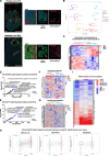
Fig. 1
Fig. 1
A Digital Spatial profiling experiment of 24 ROIs selected within epithelial-tumor Pan-Cytokeratin + (CK+ Vim) (green signal) and stromal Vimentin + (CKVim+) (red signal) of primary breast adenocarcinoma lesion (PT) and corresponding hepatic metastatic foci (Met). Original magnification, × 50. Scale bar, 250 μm. B Principal component projection (PCA) of the 24 regions of interests (ROIs) within primary breast cancer tumor and liver metastases based on the 457 most highly variable genes profiled by Nanostring Digital Spatial Profiling. Principal component 1 (PC1) splits the ROIs by epithelial-tumor (CK+Vim) or stromal (CKVim.+) component, while Principal component 2 (PC2) splits ROIs by PT and Met. C Hierarchical clustering based on the stromal Met/PT spatial signature. The signature discriminates Met and PT stromal ROIs. D-E Pathway enrichment of 129 stromal Met spatial signature genes and 99 stromal PT spatial signature genes (Reactome Pathway library). Significant pathways are highlighted in blue (adj-p.value < 0.05), with the names of the most significant ones labeled in the figure. F-G Expression of “Complement System” and “Lymphocyte Regulation” genes in Met and PT stromal ROIs showed significant differences between the two ROI groups. The heatmap left bar indicates the significant differentially expressed genes between Met and PT ROIs (orange). H Distributions and statistical comparison of the Met/PT spatial signature combined expression in breast cancer primary-metastasis pairs from the GEICAM trial (n = 70 pairs), from the University of North Carolina Rapid Autopsy Program dataset (RAP-study; n = 67 pairs), and Aftimos et al. AURORA dataset (n = 108 pairs). The paired t-test p-values confirm that the stromal Met/PT spatial signature genes differ significantly between metastatic and primary tumors samples, with Met genes upregulated in metastatic samples and PT genes in primary tumors. I Log-FC values from the comparison between Met vs PT in four different datasets, considering only the stromal Met/PT spatial signature genes whose differential expression is consistent across datasets. Positive log-FC values indicate genes upregulated in Met (red cells in the heatmap), while negative log-FC values indicate genes downregulated in Met (blue cells in the heatmap)
Fig. 2
Fig. 2
A Graphical abstract of the in vivo 4T1 triple-negative breast cancer cells injection experiment. B Representative microphotographs of H&E-stained FFPE sections from high and low lung metastatic burden involved in the Visium spatial transcriptome experiment profiling. Original magnification, × 50. Scale bar, 250 μm. C Unsupervised clustering of spatial microregions. D-E UMAP and Spatial projections highlighting foci microregions in metastasis samples. The foci microregions significantly overlap with cluster 5 (p-value < 10–16). F-G UMAP and Spatial projections of the stromal Met spatial signature total expression in metastasis samples. The Met spatial signature is over-expressed in the foci microregions. H Prediction of the foci microregions based on the stromal Met spatial signature expression. I Kaplan–Meier analysis representing the probability of survival in breast cancer patients from the BC compendium and the METABRIC dataset stratified as low Met—high PT (n = 1036 and n = 217 in the BC compendium and METABRIC, respectively) and high Met—low PT (n = 957 and n = 218 in the BC compendium and METABRIC, respectively) spatial signature score. The log-rank test P-value reflects the significance of the association between levels of the Met/PT spatial signature score and shorter survival

References

    1. Valastyan S, Weinberg RA. Tumor metastasis: molecular insights and evolving paradigms. Cell. 2011;147:275–92. - PMC - PubMed
    1. Alečković M, McAllister SS, Polyak K. Metastasis as a systemic disease: molecular insights and clinical implications. Biochim Biophys Acta Rev Cancer. 2019;1872(1):89–102. 10.1016/j.bbcan.2019.06.002. Epub 2019 Jun 14. PMID: 31202687; PMCID: PMC6692219. - PMC - PubMed
    1. Zhan Q, Liu B, Situ X, Luo Y, Fu T, Wang Y, Xie Z, Ren L, Zhu Y, He W, Ke Z. New insights into the correlations between circulating tumor cells and targetvorgan metastasis. Signal Transduct Target Ther. 2023;8(1):465. 10.1038/s41392-023-01725-9. PMID: 38129401; PMCID: PMC10739776. - PMC - PubMed
    1. Peinado H, et al. Pre-metastatic niches: organ-specific homes for metastases. Nat Rev Cancer. 2017;17:302–17. - PubMed
    1. Steeg PS. Targeting metastasis. Nat Rev Cancer. 2016;16:201–18. - PMC - PubMed

Substances