

Development of a deep learning-based model for guiding a dissection during robotic breast surgery
- PMID: 40065440
- PMCID: PMC11895239
- DOI: 10.1186/s13058-025-01981-3
Development of a deep learning-based model for guiding a dissection during robotic breast surgery
Abstract
Background: Traditional surgical education is based on observation and assistance in surgical practice. Recently introduced deep learning (DL) techniques enable the recognition of the surgical view and automatic identification of surgical landmarks. However, there was no previous studies have conducted to develop surgical guide for robotic breast surgery. To develop a DL model for guiding the dissection plane during robotic mastectomy for beginners and trainees.
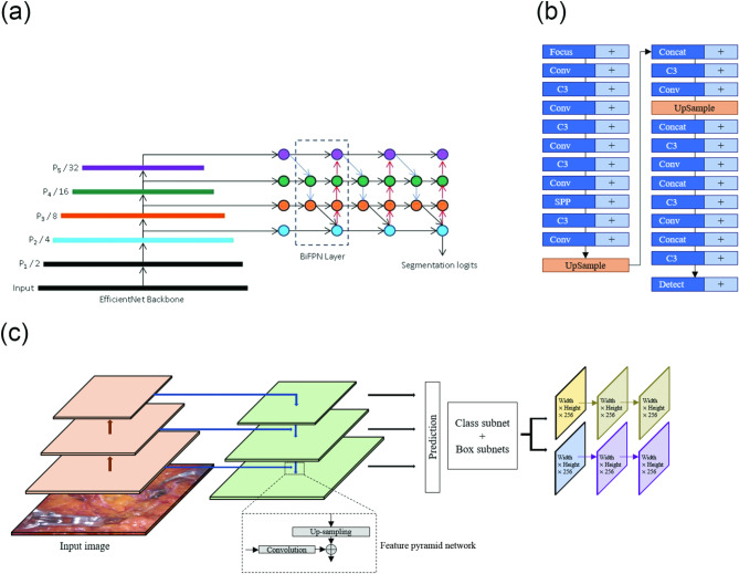
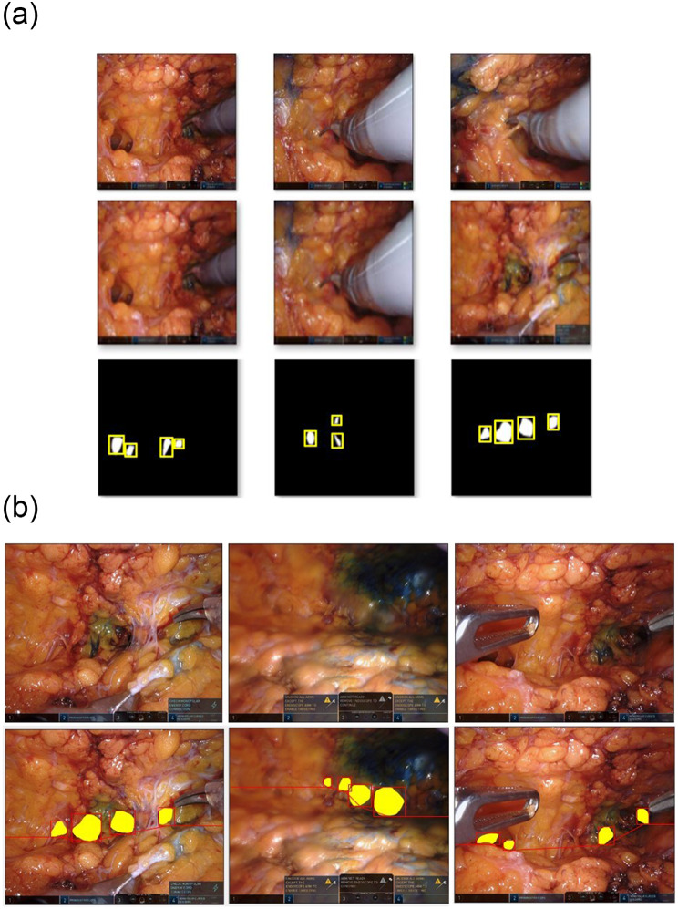
Methods: Ten surgical videos of robotic mastectomy procedures were recorded. Video frames taken at 1-s intervals were converted to PNG format. The ground truth was manually delineated by two experienced surgeons using ImageJ software. The evaluation metrics were the Dice similarity coefficient (DSC) and Hausdorff distance (HD).
Results: A total of 8,834 images were extracted from ten surgical videos of robotic mastectomies performed between 2016 and 2020. Skin flap dissection during the robotic mastectomy console time was recorded. The median age and body mass index of the patients was 47.5 (38-52) years and 22.00 (19.30-29.52) kg/m2, respectively, and the median console time was 32 (21-48) min. Among the 8,834 images, 428 were selected and divided into training, validation, and testing datasets at a ratio of 7:1:2. Two experts determined that the DSC of our model was 0.828[Formula: see text]5.28 and 0.818[Formula: see text]6.96, while the HDs were 9.80[Formula: see text]2.57 and 10.32[Formula: see text]1.09.
Conclusion: DL can serve as a surgical guide for beginners and trainees, and can be used as a training tool to enhance surgeons' surgical skills.
Keywords: Artificial intelligence; Breast neoplasms; Deep learning; Minimally invasive surgical procedures; Robotic mastectomy.
© 2025. The Author(s).
Conflict of interest statement
Declarations. Ethical approval: This study was approved by Severance Hospital Institutional Review Board (approval no. 4-2021-0241). Written informed consent was obtained from the patient for publication of accompanying images videos. A copy of the written consent is available for review by the Editor-in-Chief of this journal on request. Competing of interests: The authors declare no competing interests.
Figures


Similar articles
-
Utilization of artificial intelligence in minimally invasive right adrenalectomy: recognition of anatomical landmarks with deep learning.Acta Chir Belg. 2024 Dec;124(6):492-498. doi: 10.1080/00015458.2024.2363599. Epub 2024 Jun 10. Acta Chir Belg. 2024. PMID: 38841838
-
Breast cancer robotic nipple sparing mastectomy: evaluation of several surgical procedures and learning curve.World J Surg Oncol. 2019 Feb 6;17(1):27. doi: 10.1186/s12957-019-1567-y. World J Surg Oncol. 2019. PMID: 30728011 Free PMC article.
-
An artificial intelligence-based nerve recognition model is useful as surgical support technology and as an educational tool in laparoscopic and robot-assisted rectal cancer surgery.Surg Endosc. 2024 Sep;38(9):5394-5404. doi: 10.1007/s00464-024-10939-z. Epub 2024 Jul 29. Surg Endosc. 2024. PMID: 39073558 Free PMC article.
-
Postoperative complications and surgical outcomes of robotic versus conventional nipple-sparing mastectomy in breast cancer: meta-analysis.Br J Surg. 2024 Jan 3;111(1):znad336. doi: 10.1093/bjs/znad336. Br J Surg. 2024. PMID: 37890072 Free PMC article.
-
Use of Magnetic Resonance Imaging for Evaluating Residual Breast Tissue After Robotic-Assisted Nipple-Sparing Mastectomy in Women With Early Breast Cancer.Korean J Radiol. 2023 Jul;24(7):640-646. doi: 10.3348/kjr.2022.0708. Korean J Radiol. 2023. PMID: 37404106 Free PMC article. Review.
References
-
- Giger ML. Machine learning in Medical Imaging. J Am Coll Radiol. 2018;15(3 Pt B):512–20. - PubMed
MeSH terms
Grants and funding
LinkOut - more resources
Full Text Sources
Medical
