. 2025 Jul 1;117(7):1387-1400.
doi: 10.1093/jnci/djaf060.

Determinants of late metastases in renal cell carcinoma

Affiliations

Determinants of late metastases in renal cell carcinoma

Payal Kapur et al. J Natl Cancer Inst. .

Abstract

Background: The mechanisms underlying metastatic latency in renal cell carcinoma (RCC) remain poorly understood.

Methods: This study evaluated 2 large independent cohorts for differences in tumor biology between patients who developed metastases early (≤1 year after nephrectomy) and those with late onset (>3 years).

Results: In the discovery cohort (n = 161), late metastatic RCC was associated with clear cell histology (88.9% vs 78.7%), lower pathological stage (pT1-2; 40.3% vs 18.0%), and favorable histopathological features including low grade (40.0% vs 2.3%), less sarcomatoid (5.6% vs 21.8%), and reduced necrosis (37.7% vs 78.3%; all P < .02). Late metastatic RCC tumors exhibited increased angiogenesis (63.5% vs 19.4%) and reduced inflammation (78.8% vs 50.0%; all P < .02) profiles. Genomic driver analyses revealed comparable rates of PBRM1 and SETD2 loss in late and early metastatic RCC, while BAP1 loss was significantly less common in late metastatic RCC (7.5% vs 27.1%; P < .02). In multivariable models, BAP1/PBRM1/SETD2 status and tumor necrosis emerged as key discriminators of late metastatic RCCs. These findings were confirmed in the second cohort (n = 307). Late metastatic RCC was enriched for fatty acid oxidation and angiogenesis pathways, supporting a less aggressive phenotype. This was further evidenced by a lower engraftment rate in murine models (0% vs 36.5%; P < .001) and significantly longer overall survival from the time of metastasis (median survival doubled, P < .001). Interestingly, late metastatic RCC shared genomic and phenotypic features with RCC that metastasizes to the pancreas, suggesting a common underlying biology influencing both metastatic latency and pancreatic tropism.

Conclusions: Overall, these findings advocate for recognition of late metastatic RCC because of its distinct biology and improved prognosis.


Conflict of interest statement

No relevant conflicts of interest.

Figures
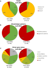
Figure 1.
Figure 1.
Late metastatic renal cell carcinomas are characterized by indolent histology and molecular features. Integrated pie charts of (A) grade (UTSW and Mayo Clinic cohorts), (B) dominant architectural patterns (clear cell renal cell carcinoma, UTSW cohort), and (C) driver gene assessed by immunohistochemical analyses of the corresponding protein (or mark) in (clear cell renal cell carcinoma, UTSW and Mayo Clinic cohorts) for primary tumors in late metastatic renal cell carcinoma vs early metastatic renal cell carcinoma (see tables for individual cohort data). Patients with missing data for any of the 3 immunohistochemistries were excluded in the immunohistochemistry pie chart. Loss of BAP1 (irrespective of other gene status) was categorized as BAP1 loss. Abbreviations: – = protein not detected by immunohistochemistry; + = protein detected by immunohistochemitry; mRCC = metastatic renal cell carcinoma; UTSW = University of Texas Southwestern Medical Center.
Figure 3.
Figure 3.
Differential engraftment rates of late metastatic renal cell carcinoma and early metastatic renal cell carcinoma in mice. Pie charts illustrating engraftment rates of corresponding primary tumors in NOD/SCID mice. Abbreviation: mRCC = metastatic renal cell carcinoma.
Figure 2.
Figure 2.
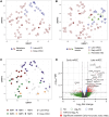
Gene expression analyses of late metastatic renal cell carcinoma from clear cell renal cell carcinoma. A) Uniform manifold approximation and projection analyses of whole transcriptome from clear cell renal cell carcinomas that developed late and early metastases. B) Uniform manifold approximation and projection merged with an additional cohort of clear cell renal cell carcinoma with pancreatic metastases. C) Uniform manifold approximation and projection (from Figure 2A) with overlayed nonnegative matrix factorization cluster designation. D) Volcano plot of differentially expressed genes in early metastatic renal cell carcinoma and late metastatic renal cell carcinoma/pancreatic metastases. Abbreviations: btw = between; ccRCC = clear cell renal cell carcinoma; FC = fold-change; FDR = significant after false discovery rate; Log2FC = significant based on gene expression fold change; mRCC = metastatic renal cell carcinoma; mets = metastasis; NMF = nonnegative matrix factorization; NS = not significant; UMAP = uniform manifold approximation and projection.
Figure 4.
Figure 4.
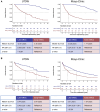
Kaplan–Meier analyses of late metastatic renal cell carcinoma and early metastatic renal cell carcinoma patients. Kaplan–Meier analyses of late metastatic renal cell carcinoma patients compared with early metastatic renal cell carcinoma for (A) overall survival from time of nephrectomy and (B) overall survival from time of metastases diagnosis, for University of Texas Southwestern Medical Center and Mayo Clinic cohorts. Abbreviations: CI = confidence interval; HR = hazard ratio; mRCC = metastatic renal cell carcinoma; UTSW = University of Texas Southwestern Medical Center.

References

    1. Siegel RL, Miller KD, Jemal A. Cancer statistics, 2020. CA Cancer J Clin. 2020;70:7-30. 10.3322/caac.21590 - DOI - PubMed
    1. Siegel RL, Miller KD, Fuchs HE, Jemal A. Cancer statistics, 2022. CA Cancer J Clin. 2022;72:7-33. 10.3322/caac.21708 - DOI - PubMed
    1. Dabestani S, Thorstenson A, Lindblad P, Harmenberg U, Ljungberg B, Lundstam S. Renal cell carcinoma recurrences and metastases in primary non-metastatic patients: a population-based study. World J Urol. 2016;34:1081-1086. 10.1007/s00345-016-1773-y - DOI - PubMed
    1. Padala SA, Barsouk A, Thandra KC, et al. Epidemiology of renal cell carcinoma. World J Oncol. 2020;11:79-87. 10.14740/wjon1279 - DOI - PMC - PubMed
    1. Zisman A, Pantuck AJ, Dorey F, et al. Improved prognostication of renal cell carcinoma using an integrated staging system. Validation Studies. J Clin Oncol. 2001;19:1649-1657. 10.1200/JCO.2001.19.6.1649 - DOI - PubMed

MeSH terms