Review
. 2025;2(1):11.
doi: 10.1038/s44323-025-00025-5. Epub 2025 Mar 8.

Dual orexin receptor antagonists as promising therapeutics for Alzheimer's disease

Affiliations
Review

Dual orexin receptor antagonists as promising therapeutics for Alzheimer's disease

S M Ragsdale et al. NPJ Biol Timing Sleep. 2025.

Abstract

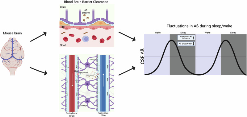
We examine the relationship between sleep, glymphatics and Alzheimer's disease (AD), and recent work questioning glymphatic clearance during sleep. We highlight a need for understanding glymphatic and/or other mechanism of clearance during sleep, and review glymphatic flow measurement methods. Further, we explore dual orexin receptor antagonists (DORAs) potential to mitigate AD sleep disturbances and enhance clearance. Further research could elucidate a linkage between DORAs, improved sleep and reducing AD pathophysiology.

Keywords: Biological techniques; Diseases; Neuroscience.


Conflict of interest statement

Competing interestsCompeting interests from W.V.M. include research support from Axon Medical Technologies and scientific advising from Haleon, Idorsia, LivaNova, and Carelon.

Figures

Fig. 1
Fig. 1. In the mouse, there is a relationship between brain waste clearance and fluctuations in levels of amyloid beta in the brain during sleep/wake.
The mechanism that facilitates these fluctuations may be through blood-brain barrier (BBB) clearance or glymphatic clearance, both of which contribute to clearance during sleep. Understanding the mechanisms that aid in reducing amyloid beta levels during sleep is important for understanding Alzheimer’s disease (AD) pathology. Note, the recent finding that glymphatic flow may be lower during sleep does not account for the Aquaphorin-4 APQ4 literature, and APQ4 deletion does not impact BBB clearance. Therefore, it remains that the mechanism of amyloid clearance during sleep is likely at least partially mediated by the glymphatic system (see text for details). This figure was generated using BioRender.
Fig. 2
Fig. 2. Graphical summary of methods for assessing glymphatic flow with key references.
Non-imaging methods to evaluate glymphatic flow include CSF and plasma assays, which provide direct concentration measurements of the products of interest but are invasive. The imaging methods utilize various contrast agents and tracers, which can be tuned by size to interrogate different aspects of the glymphatic system. Fluorescence microscopy was able to define the glymphatic system initially and has high resolution and specificity; however, it has a limited field of view and cannot probe deep into the brain. PET can provide dynamic information, is highly sensitive and selective, but has limited resolution. MRI can use endogenous contrast in addition to exogenous agents, making it less invasive and provides a wider range of imaging capabilities, while also being highly translatable to clinical work. This figure was generated using BioRender.
Fig. 3
Fig. 3. Sleep reduces TAβA potentially by glymphatic clearance.
Top. Depiction of hypothetical CSF Aβ fluctuations in a mouse with Aβ and Tau aggregation. AD pathology is thought to lead to lower glymphatic clearance during sleep/wake cycles and as a consequence higher Aβ production. Bottom. Regardless of the exact mechanism, if clearance is occurring during sleep, then improving sleep quality to reduce fragmented sleep may be one strategy to slow AD further Aβ and Tau aggregation. Treating AD mice with Dual Orexin Receptor Antagonists (DORAs) could potentially regulate Aβ fluctuations during sleep/wake cycles by increasing glymphatic clearance and lowering net Aβ production. This figure was generated using BioRender.

References

    1. Poddar, M. K. et al. Metabolic disorder in Alzheimer’s disease. Metab. Brain Dis.36, 781–813 (2021). - PubMed
    1. Tzioras, M. et al. Synaptic degeneration in Alzheimer disease. Nat. Rev. Neurol.19, 19–38 (2023). - PubMed
    1. Lee, S. J. et al. Protein aggregate spreading in neurodegenerative diseases: problems and perspectives. Neurosci. Res70, 339–348 (2011). - PMC - PubMed
    1. Bisaz, R., Travaglia, A. & Alberini, C. M. The neurobiological bases of memory formation: from physiological conditions to psychopathology. Psychopathology47, 347–356 (2014). - PMC - PubMed
    1. Cushing, S. D. et al. Rescuing impaired hippocampal-cortical interactions and spatial reorientation learning and memory during sleep in a mouse model of Alzheimer’s disease using hippocampal 40 Hz stimulation. 10.1101/2024.06.20.599921 (2024).

LinkOut - more resources