. 2025 Mar 10:6:0117.
doi: 10.34133/cbsystems.0117. eCollection 2025.

Application of Ultrasound Localization Microscopy in Evaluating the Type 2 Diabetes Progression

Affiliations

Application of Ultrasound Localization Microscopy in Evaluating the Type 2 Diabetes Progression

Tao Zhang et al. Cyborg Bionic Syst. .

Abstract

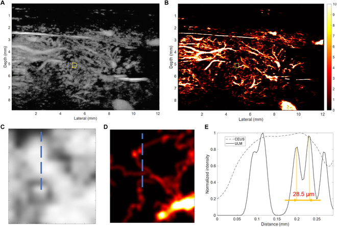
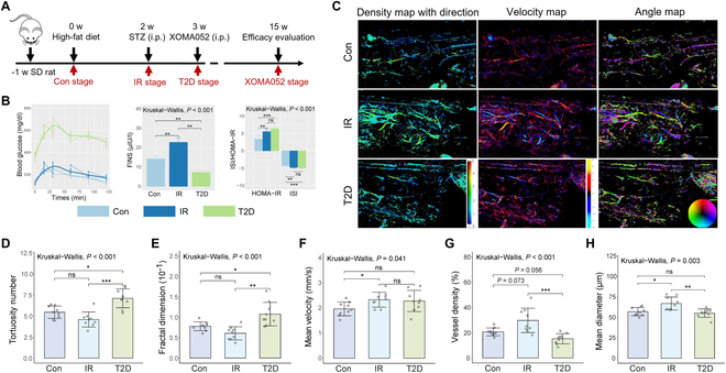
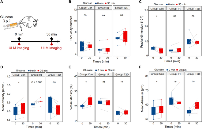
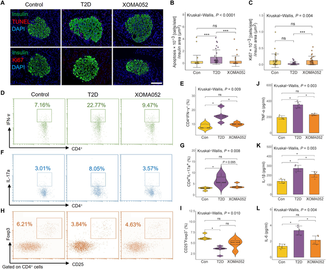
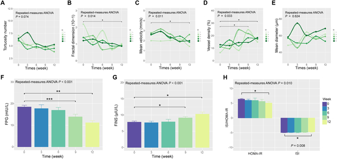
Type 2 diabetes is considered as a chronic inflammatory disease in which the dense microvasculature reorganizes with disease progression and is highly correlated with β cell mass and islet function. In this study, we constructed rat models of type 2 diabetes and used ultrasound localization microscopy (ULM) imaging to noninvasively map the pancreatic microvasculature at microscopy resolution in vivo to reflect β cell loss and islet function deterioration, and evaluate the efficacy after anti-cytokine immunotherapy. It was unveiled that ULM morphological and hemodynamic parameters have a strong link with β cell loss and deterioration of pancreatic islet function. This correlation aligns with the observed pathological alterations in the microvessels of islet and demonstrated that ULM can effectively mirror the functionality of β cells during rapid fluctuations in blood glucose levels by observing changes in mean velocity. Furthermore, it was revealed that treatment with anti-cytokine immunotherapy enhances the function and health of β cells by restoring the microvascular environment. Remarkable improvements in vessel morphology (measured by fractal dimension) and hemodynamics (indicated by mean velocity and vessel density) were noted following the anti-cytokine immunotherapy, signifying a significant enhancement at the treatment's conclusion (P < 0.05). These observations suggested that ULM technology holds promise as a visible and efficient tool for monitoring the effectiveness of anti-cytokine immunotherapy in managing type 2 diabetes. Pancreatic microvessel-based ULM may serve as a novel noninvasive method to assess β cells, providing a valuable clinical tool for tracking the progression of type 2 diabetes.


Conflict of interest statement

Competing interests: The authors declare that they have no competing interests.

Figures

Fig. 1.
Fig. 1.
Diagram of ULM processing.
Fig. 2.
Fig. 2.
The resolution improvement and quantification of the ULM image. (A) CEUS MIP image and (B) ULM density image of pancreas. (C) Magnified CEUS MIP and (D) ULM density images of the same region within the yellow boxes. (E) Vessel intensity profiles along the blue line on the magnified CEUS MIP and ULM images.
Fig. 3.
Fig. 3.
Longitudinal study of real-time ULM imaging in vivo using a type 2 diabetes model. (A) Study design. (B) The intraperitoneal glucose tolerance test (ipGTT), fasting serum insulin (FINS), homeostatic model assessment of insulin resistance (HOMA-IR) and insulin resistance index (ISI) of T2D progression. (C) ULM imaging in the progression of T2D, including density map with direction, velocity map, and angle map. ULM values in the progression of T2D, (D) tortuosity number, (E) fractal dimension, (F) mean velocity, (G) vessel density and (H) mean diameter. (n = 9). * = P < 0.05, ** = P < 0.01, *** = P < 0.001, ns = nonsignificant.
Fig. 4.
Fig. 4.
Changes in islet microvascular morphology in SD rats. (A) Representative maximum-projection confocal image between 8- to 10-μm depth of pancreas section from SD rat infused with FITC-conjugated tomato insulin (green), lectin (red), and DAPI (blue) immunofluorescence staining of pancreatic islets in different stages of T2D. Scale bar, 20 µm. (B) Vascular diameter in different disease stages of T2D. (C) Vascular coverage in islet and (D) in exocrine tissue in the T2D progression (n = 25 rat islets from 5 rats). (E) β cell volume and (F) β cell mass in different disease stages of T2D (n = 15 panoramic scans of pancreas from 5 rats). *P < 0.05, **P < 0.01, ***P < 0.001, ns = nonsignificant.
Fig. 5.
Fig. 5.
Noninvasive β cell function evaluation in vivo after glucose stimulation. (A) Study design. Measurements of (B) tortuosity number, (C) fractal dimension, (D) mean velocity, (E) vessel density, and (F) mean diameter in the control, IR, and T2D groups (n = 5). *P < 0.05, ns = nonsignificant.
Fig. 6.
Fig. 6.
XOMA052 promotes β cell proliferation and evaluates the efficacy of anti-cytokine immunotherapy. (A) Fluorescence staining image of TUNEL and Ki67 of pancreatic islets in SD rats. Scaler bar, 100 μm. Corresponding positive cell number of (B) TUNEL and (C) Ki67 in fluorescence staining image (n = 30 rat islets from 5 rats). (D and E) Representative flow cytometry plots showing the CD4+ IFN-ɤ+ T cells in pancreatic islets and quantification. (F and G) Representative flow cytometry plots showing the CD4+ IL-17a+ T cells in pancreatic islets and quantification. (H and I) Representative flow cytometry plots showing the CD4+ CD25+ Foxp3+ T cells in pancreatic islets and quantification. Cytokine levels of (J) TNF-α, (K) IL-1β, and (L) IL-6 in sera from rats in different stages (n = 5). *P < 0.05, ***P < 0.001, ns = nonsignificant.
Fig. 7.
Fig. 7.
ULM imaging in efficacy evaluation of anti-cytokine immunotherapy in type 2 diabetes. (A) The tortuosity number, (B) fractal dimension, (C) mean velocity, (D) vessel density, (E) mean diameter, (F) fast plasma glucose (FPG), (G) fasting serum insulin (FINS), (H) homeostatic model assessment of insulin resistance (HOMA-IR), and insulin resistance index (ISI) of T2D rats evaluated at 0, 3, 6, 9, and 12 weeks after anti-inflammatory treatment with XOMA052 (n = 5). *P < 0.05, **P < 0.01, ***P < 0.001.

References

    1. Donath MY, Shoelson SE. Type 2 diabetes as an inflammatory disease. Nat Rev Immunol. 2011;11(2):98–107. - PubMed
    1. Rohm TV, Meier DT, Olefsky JM, Donath MY. Inflammation in obesity, diabetes, and related disorders. Immunity. 2022;55(1):31–55. - PMC - PubMed
    1. He H, Fasoula NA, Karlas A, Omar M, Aguirre J, Lutz J, Kallmayer M, Füchtenbusch M, Eckstein HH, Ziegler A, et al. . Opening a window to skin biomarkers for diabetes stage with optoacoustic mesoscopy. Light Sci Appl. 2023;12(1):231. - PMC - PubMed
    1. Yang J, Zhang LJ, Wang F, Hong T, Liu Z. Molecular imaging of diabetes and diabetic complications: Beyond pancreatic beta-cell targeting. Adv Drug Deliv Rev. 2019;139:32–50. - PubMed
    1. Siehler J, Blochinger AK, Meier M, Lickert H. Engineering islets from stem cells for advanced therapies of diabetes. Nat Rev Drug Discov. 2021;20(12):920–940. - PubMed

LinkOut - more resources