. 2025 Feb 24:16:1544154.
doi: 10.3389/fimmu.2025.1544154. eCollection 2025.

Unveiling SSR4: a promising biomarker in esophageal squamous cell carcinoma

Affiliations

Unveiling SSR4: a promising biomarker in esophageal squamous cell carcinoma

Jiaqi Zhang et al. Front Immunol. .

Abstract

Background: Esophageal squamous cell carcinoma (ESCC) represents a frequent cancer with a poor prognosis. Altered glucose metabolism contributes factor to ESCC progression. In our previous study, signal sequence receptor subunit delta (SSR4) was included in an ESCC prognostic model; however, the mechanisms underlying SSR4 implication in ESCC remain ambiguous. Accordingly, we aim to determine the interconnection between SSR4 expression and clinical characteristics of ESCC.

Methods: This differential expression and prognostic significance of SSR4 was performed using bulk RNA-seq data and 110 patients with complete follow-up information. The ESCC cell subsets with the highest gene expression levels were identified with single-cell data. Gene function and enrichment, immune infiltration, cell communication, and molecular docking analyses were performed.

Results: Unlike adjacent non-cancerous tissues, SSR4 was overexpressed in ESCC tissues, validated by both reverse transcription-qPCR and IHC staining. SSR4 expression was related to the N stage, lymph node metastasis, and AJCC TNM classification stage. Patients exhibiting low SSR4 expression had a more favorable prognosis. The highest SSR4 expression was recognized in tumor plasma cells. Continued exploration of immune infiltration highlighted a close association between SSR4 gene expression and the infiltration of immune cells such as plasma cells. On dividing cells into SSR4-positive and -negative groups, CellChat analysis indicated that SSR4 may regulate the interactions that existed between ESCC tumor plasma cells and the tumor microenvironment (TME) by modulating the MIF/CD74/CXCR4 axis.

Conclusion: The SSR4 gene may have significant relevance with clinical pathological factors, and play a critical role in the regulation of tumor microenvironment of ESCC patients. Overall, SSR4 may be a promising ESCC biomarker with prospective applicability in clinical diagnosis as well as the development of targeted treatment approaches in patients of ESCC.

Keywords: SSR4; biomarker; esophageal squamous cell carcinoma; immune cells; single-cell transcriptomics.


Conflict of interest statement

The authors declare that the research was conducted in the absence of any commercial or financial relationships that could be construed as a potential conflict of interest.

Figures

Figure 1
Figure 1
Study flowchart. ESCC, esophageal squamous cell carcinoma; SSR4, signal sequence receptor subunit delta; DEGs, differentially expressed genes.
Figure 2
Figure 2
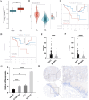
Signal sequence receptor subunit delta (SSR4) identification in esophageal squamous cell carcinoma (ESCC) patients. (A) The Cancer Genome Atlas (TCGA) database: Difference expression of SSR4 in ESCA and para-cancerous (P < 0.001). (B) TCGA and Genotype-Tissue Expression (GTEx) databases: Difference in expression of SSR4 in ESCC and healthy individuals (P < 0.0001). (C) High SSR4 expression was related to poor overall survival (P = 0.016) and (D) poor disease-free survival in ESCC patients (P = 0.0286). (E) SSR4 numbers in ESCC tissues and adjacent tissues (P < 0.0001) and (F) matched para-cancerous tissues (P < 0.0001). (G) Results of RT-qPCR demonstrated that SSR4 was highly expressed in ESCC cell lines. Analysis was performed on data from three independent experiments. (H) Tissue microarray and immunohistochemistry staining of SSR4 in ESCC and adjacent tissue (magnification x10 and x20; scale bar: 100 and 50 µm, respectively). Notes: DEGs, differentially expressed genes; ESCA, esophageal carcinoma. (**P < 0.01, ***P < 0.001, ****P < 0.0001.).
Figure 3
Figure 3
Clustering and annotation of the ESCC cells. (A, B) Sixteen clusters were annotated into 10 cell types. (C) The bubble chart shows specific markers corresponding to 10 different cell types. (D) Variations in the content percentages of 10 cell types in 8 groups of samples. (E) Cell distributions differ between individual samples in control and disease groups.
Figure 4
Figure 4
Assessing the possible implication of signal sequence receptor subunit delta (SSR4) at a single-cell level and immune infiltration. (A, B) Different cell types’ distribution and expression of SSR4. (C) Correlation between SSR4 and immune infiltrating cells based on ssGSEA algorithm. (D, E) Two groups of plasma cells were determined based on their expression of SSR4: SSR4+ and SSR4-. (*P < 0.05, **P < 0.01.).
Figure 5
Figure 5
Single-gene functional enrichment analysis. (A) Analysis of functional enrichment in GO and (B) KEGG: Barplot. (C) SSR4+ and SSR4- expressions correlate with metabolic pathways.
Figure 6
Figure 6
Cell communication suggests that signal sequence receptor subunit delta (SSR4) may affect tumor progression in ESCC via macrophage migration inhibitory factor (MIF) signaling. (A) Diagram depicting interaction signal frequency and strength among different cells. (B) Plasma-SSR4+ and -SSR4- cells emit different signals. (C) A heatmap: Interaction signal intensity between diverse cell types. (D) Cells of the Plasma-SSR4+ and -SSR4- lineages interact with each other through the MIF signaling pathway. (E) Bubble diagram: Difference between Plasma-SSR4+ and -SSR4- cells.

References

    1. Anandavadivelan P, Lagergren P. Cachexia in patients with esophageal cancer. Nat Rev Clin Oncol. (2016) 13:185–98. doi: 10.1038/nrclinonc.2015.200 - DOI - PubMed
    1. Sung H, Ferlay J, Siegel RL, Laversanne M, Soerjomataram I, Jemal A, et al. . Global cancer statistics 2020: GLOBOCAN estimates of incidence and mortality worldwide for 36 cancers in 185 countries. CA Cancer J Clin. (2021) 71:209–49. doi: 10.3322/caac.21660 - DOI - PubMed
    1. Watanabe M, Otake R, Kozuki R, Toihata T, Takahashi K, Okamura A, et al. . Recent progress in multidisciplinary treatment for patients with esophageal cancer. Surg Today. (2020) 50:12–20. doi: 10.1007/s00595-019-01878-7 - DOI - PMC - PubMed
    1. Bolidong D, Domoto T, Uehara M, Sabit H, Okumura T, Endo Y, et al. . Potential therapeutic effect of targeting glycogen synthase kinase 3β in esophageal squamous cell carcinoma. Sci Rep. (2020) 10:11807. doi: 10.1038/s41598-020-68713-9 - DOI - PMC - PubMed
    1. Puhr HC, Preusser M, Ilhan-Mutlu A. Immunotherapy for esophageal cancers: what is practice changing in 2021? Cancers (Basel). (2021) 13(18):4632. doi: 10.3390/cancers13184632 - DOI - PMC - PubMed

MeSH terms

Substances

LinkOut - more resources