. 2025 Feb 27;53(5):gkaf169.
doi: 10.1093/nar/gkaf169.

Amyloid-like DNA bridging: a new mode of DNA shaping

Affiliations

Amyloid-like DNA bridging: a new mode of DNA shaping

Frank Wien et al. Nucleic Acids Res. .

Abstract

All organisms depend on specific proteins to compact and organize their genomes. In eukaryotes, histones fulfil this role, while bacterial chromosomes are shaped by nucleoid-associated proteins (NAPs). Among its pleiotropic functions, the NAP Hfq plays a pivotal role in bacterial genome organization. In this study, we characterized the structure of the C-terminal extension of Hfq, which mediates chromosomal compaction, in its DNA-bound state. Using an integrative approach that combined transmission electron microscopy, neutron scattering, site-directed mutagenesis, and molecular modeling, we identified an amyloid module formed by the C-terminal region of Hfq. This module uniquely bridges and compacts six DNA molecules, marking the first documented instance of an amyloid structure with DNA-bridging properties. Our findings redefine the functional landscape of amyloids, linking them to genome architecture and gene regulation. This result suggests that amyloid-DNA interactions may represent a conserved mechanism across biological systems, with profound implications for understanding genome organization and the regulation of gene expression in both prokaryotes and eukaryotes.


Conflict of interest statement

The authors declare no conflict of interest.

Figures

Graphical Abstract
Graphical Abstract
Figure 1.
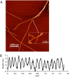
Figure 1.
AFM images of fibrillar Hfq-CTR:DNA complexes on mica. (A) Fibers adopting a helical structure. Note the tendency of the fibers to intertwine at the top of the panel. The inset shows a tilted view of long fibers, revealing 2–3 nm protrusions spaced by ∼100 nm. (B) The profile of the top of the fiber, taken from the region indicated by the two white arrows.
Figure 2.
Figure 2.
Cryo-EM image of the Hfq-CTR:DNA fibers. Representative aligned micrograph from a set of 1100 movies acquired. Arrows indicate the two kinds of fibers found in this work: thin fibers (10–15 nm diameter) and thicker fibers (25 nm diameter). The scale bar represents 50 nm.
Figure 3.
Figure 3.
Asymmetrical helical reconstruction of the fiber using SPA. (A) Close-up view of a section in the thinner part of 3D reconstruction, as shown in panel (B). (C) Symmetry analysis of the 3D reconstructed fiber. A 6-fold symmetry is observed for the reconstructed fiber, with slight outward deformation confirming the right-handed helical nature of the fiber.
Figure 4.
Figure 4.
SANS analysis. (A) Cross-sectional form factor for the DNA component of the fiber. The solid curve represents the fit of the form factor expression. (B) Two-dimensional DNA density profile of the fiber’s cross-section. (C) Azimuthally averaged radial DNA density profile. (DF) As in panels (A–C) but for the Hfq-CTR component.
Figure 5.
Figure 5.
INS spectra. Spectra were collected at 10 K all in D2O for deuterated crystalline ice Ih (gray), DNA (cyan), Hfq-CTR (blue), and Hfq-CTR:DNA complex (red), normalized to the elastic line to account for differences in density and instrument conditions. Here one should consider possible shifts due to harmonic, isotope-independent force constants, with a factor of √ (20/18) = 1.054 for the translational modes. (A) No exchange with D2O occurs, as indicated by the absence of a vibrational mode ∼900 cm−1 [50]. The softening of the 400 cm−1 vibration for the complexes, highlighted in yellow, indicates that hydration water remains undisturbed. (B) Lattice vibrations. The analysis of the overall spectral weight (or intensity) allows the conclusion that the fiber in D2O is the stiffest. (C) Spectral changes suggest the partial opening of the dsDNA and an effect of the Hfq-CTR on the deoxyribose pucker, as described in the text. (D) The strong vibrational mode at 1625 cm−1 demonstrates interactions between Hfq-CTR and the N6 amino group of adenines. An interaction at the N6 position of adenine is typically indicative of major groove recognition [57]. This major groove interaction is further supported by the fact that A-tract DNA has a compressed, narrow minor groove, which is unlikely to accommodate an amyloid structure.
Figure 6.
Figure 6.
Fibrillar complexes of Hfq-CTR and DNA. (A) Approximate positions of the centers of mass of the DNA helices in the complex used in the modeling. This graph shows the cross-sectional view from the top of the complex along the fiber axis. Note that the hole at the center of panel (A) might indicate a depletion in density rather than complete absence of Hfq-CTR at this specific cross-section of the fiber (see the radial density profile of Hfq-CTR in Fig. 4F). (B) The six green DNA double-helices arranged hexagonally in this panel correspond to the positions of the six DNAs in panel (A) after refinement. The six N-terminal torus of Hfq, which are absent in this model, should decorate the fiber and extend outward from it. (C) As suggested by both Cryo-EM and INS experiments, the dsDNA can be locally separated into two single-stranded DNA regions. This specific region was also modeled in the Cryo-EM envelope.

References

    1. Kuzminov A Bacterial nucleoid is a riddle wrapped in a mystery inside an enigma. J Bacteriol. 2024; 206:e0021123. 10.1128/jb.00211-23. - DOI - PMC - PubMed
    1. Monk JM, Koza A, Campodonico MA et al. Multi-omics quantification of species variation of Escherichia coli links molecular features with strain phenotypes. Cell Syst. 2016; 3:238–51. 10.1016/j.cels.2016.08.013. - DOI - PMC - PubMed
    1. Dorman CJ Function of nucleoid-associated proteins in chromosome structuring and transcriptional regulation. J Mol Microbiol Biotechnol. 2014; 24:316–31. - PubMed
    1. Dame RT, Goosen N HU: promoting or counteracting DNA compaction?. FEBS Lett. 2002; 529:151–6. 10.1016/S0014-5793(02)03363-X. - DOI - PubMed
    1. Owen-Hughes TA, Pavitt GD, Santos DS et al. The chromatin-associated protein H-NS interacts with curved DNA to influence DNA topology and gene expression. Cell. 1992; 71:255–65. 10.1016/0092-8674(92)90354-F. - DOI - PubMed

MeSH terms