. 2025 Apr 15;99(4):e0177724.
doi: 10.1128/jvi.01777-24. Epub 2025 Mar 11.

BET degraders reveal BRD4 disruption of 7SK and P-TEFb is critical for effective reactivation of latent HIV in CD4+ T-cells

Affiliations

BET degraders reveal BRD4 disruption of 7SK and P-TEFb is critical for effective reactivation of latent HIV in CD4+ T-cells

Anne-Marie W Turner et al. J Virol. .

Abstract

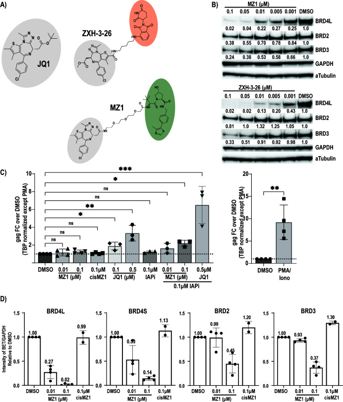
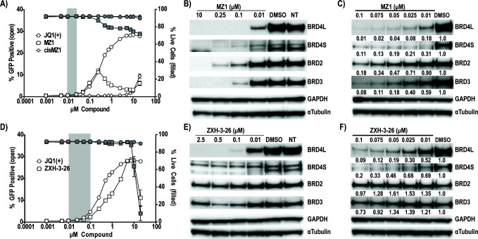
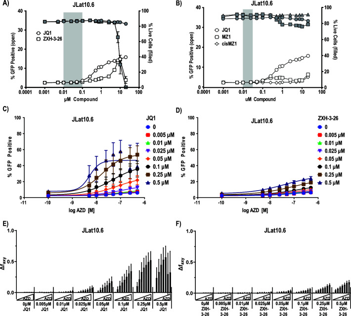
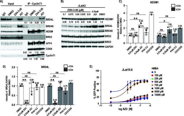
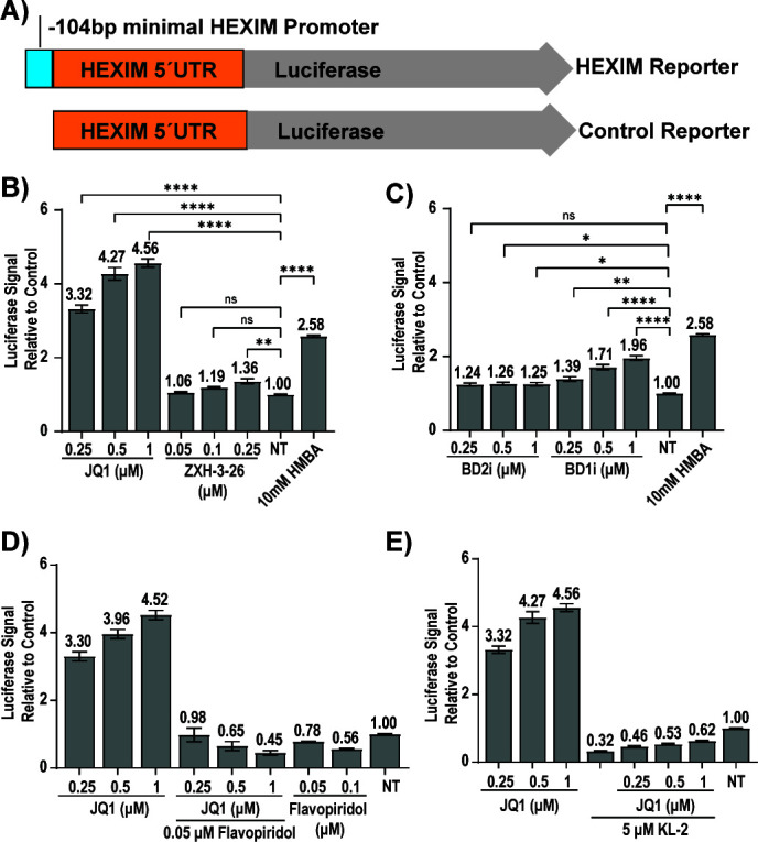
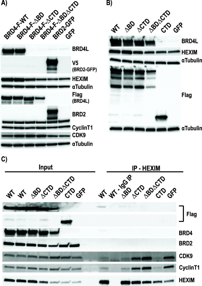
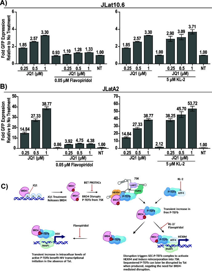
HIV cure strategies that aim to induce viral reactivation for immune clearance leverage latency reversal agents to modulate host pathways which directly or indirectly facilitate viral reactivation. Inhibition of bromo and extra-terminal domain (BET) family member BRD4 reverses HIV latency, but enthusiasm for the use of BET inhibitors in HIV cure studies is tempered by concerns over inhibition of other BET family members and dose-limiting toxicities in oncology trials. Here, we evaluated the potential for bivalent chemical degraders targeted to the BET family as alternative latency reversal agents. We observed that despite highly potent and selective BRD4 degradation in primary CD4+ T-cells from ART-suppressed donors, BRD4 degraders failed to induce latency reversal as compared to BET inhibitors. Furthermore, BRD4 degraders failed to mimic previously observed synergistic HIV reactivation between BET inhibitors and an activator of the non-canonical NF-κB pathway. Mechanistic investigation of this discrepancy revealed that latency reversal by BET inhibitors is not related to the abatement of competition between Tat and BRD4 for P-TEFb, but rather the ability of BRD4 to disrupt 7SK and increase the levels of free P-TEFb. This activity is dependent on the shift of BRD4 from chromatin-bound to soluble and retargeting of P-TEFb to chromatin, which is dependent on intact BRD4 but independent of the bromodomains.

Importance: Multiple factors and pathways contribute to the maintenance of HIV latency, including bromo and extra-terminal domain (BET) family member BRD4. While small molecule inhibitors of the BET family result in latency reversal, enthusiasm for the use of BET inhibitors in HIV cure is limited due to toxicity concerns. We examined BRD4-selective chemical degraders as alternatives to BET inhibitors but found two robust degraders failed to induce latency reversal. We observed key differences in the ability of BET inhibitors versus BET degraders to disrupt P-TEFb, a key cellular activator of transcription and a complex required for HIV reactivation. We present a new model for the role of BRD4 in HIV latency and propose that BRD4 be reconsidered as an activator rather than a repressor of HIV transcription in the context of HIV cure strategies.

Keywords: BRD4; HIV; P-TEFb; chemical degraders; latency reversal.


Conflict of interest statement

The authors declare no conflict of interest.

Figures

Fig 1
Fig 1
HIV latency reversal in ART-suppressed donors by bivalent degraders of the BET proteins. (A) BET inhibitor JQ1 is highlighted in gray. Degraders MZ1 and ZXH-3-26 use JQ1 linked to either Von Hippel-Lindau (VHL) (green) or CRBN (orange) recruiting ligands. (B) A targeted dose titration of MZ1 and ZXH-3-26 in healthy, primary CD4+ T-cells (n = 1 donor) identifies BRD4-specific degradation at 10 nM and 5 nM, respectively. Relative protein levels were determined by standardizing each protein to the GAPDH loading control, followed by expression relative to the dimethyl sulfoxide (DMSO) control. (C) Total CD4+ T-cells from ART-suppressed donors were treated for 24 hours with MZ1 alone (n = 4, SD) or in combination with Inhibitor of Apoptosis inhibitor (IAPi) AZD5582 (n = 3, SD) and induction of gag cell-associated RNA assayed by quantitative real-time PCR. Four to five replicates of 1–2E6 cells per donor per condition were assayed. Each dot represents the average of these replicates per unique donor. Phorbol 12-myristate 13-acetate (PMA)/Ionomycin results are graphed independently due to differences in normalization (see Materials and Methods). Significance was assessed relative to DMSO control by Kruskal-Wallis with uncorrected Dunn’s multiple comparison test, (*) P < 0.05, (**) P < 0.01, (***) P < 0.001, (****) P < 0.0001. (D) BET protein levels as assayed by western blot in the 4 ART-suppressed donors (n = 4, SD). Cells were treated concurrently with those for RNA isolation. BET protein levels are quantitated relative to GAPDH levels for all donors. cisMZ1 could not be assayed in all donors due to cell limitations (n = 2, range).
Fig 2
Fig 2
Latency reversal and targeted degradation of BET degraders in a Jurkat-derived Latency Model. Latency reversal was assessed via the treatment of JLatA2 cells with a 16-point dose titration of (A) MZ1 and cisMZ1. JQ1(+) data is overlayed and provided in Fig. S4. Each titration was performed two independent times with biological triplicates (n = 6, SEM) with GFP (open symbols) assessed by flow cytometry as a measure of latency reactivation and viability by live/dead stain (gray symbols). Error bars not extending past the symbol are not visible. The window of BRD4-specific degradation for MZ1 is highlighted in gray as determined by corresponding westerns for both BRD4L(ong) and BRD4S(hort) isoforms as well as BRD2 and BRD3 in JLatA2 cells (B and C). Matching experiments were performed for (D) ZXH-3-26 and the window of BRD4-selective degradation is highlighted in gray based on corresponding westerns (E and F). Relative protein levels were determined by standardizing each protein to the GAPDH loading control, followed by expression relative to the DMSO control. Western blots are representative of at least two independent experiments (also see Fig. 4B; Fig. S6B and C).
Fig 3
Fig 3
Latency reversal and synergy with AZD5582 by degrader ZXH-3-26 is attenuated in a full-length Jurkat cell line. A 16-point dose titration of (A) ZXH-3-26 and JQ1 or (B) MZ1 and JQ1 in JLat10.6 cells. Each titration was performed two independent times with biological triplicates (n = 6, SEM). Error bars not extending past the symbol are not visible. (C) 8-point dose titration of AZD-5582 and JQ1 in JLat10.6 cells. (D) 8-point dose titration of AZD-5582 and ZXH-3-26 in JLat10.6 cells. Data represents three independent experiments of each 8-point titration (n = 3, SD). Bliss synergy calculations of (E) AZD5582/JQ1 and (F) AZD5582/ZXH-3-26 in JLat10.6 cells based on data in C/D (n = 3, SD). Synergy experiments in JLatA2 cells are provided in Fig. S5.
Fig 4
Fig 4
HEXIM1 is upregulated in response to BETi but not BET PROTACs. (A) Jurkat cells were treated with vehicle control (DMSO), 0.05 µM ZXH-3-26, or 0.5 µM JQ1 for 24 hours followed by a CyclinT1 or IgG immunoprecipitation and western blot for associated proteins. Relative protein levels are provided standardized to the DMSO IP control. HEXIM1 protein levels are upregulated in (B) JLatA2 cells in response to JQ1 but not in response to ZXH-3-26. (C) This observation is repeated in both resting and total primary CD4+ T-cells in cells from healthy donors (n = 4, SD). Relative protein levels were determined by standardizing each protein to the GAPDH loading control, followed by expression relative to the DMSO control. (D) Relative BRD4L(ong) levels in treated rCD4 and tCD4 (n = 4, SD). (E) Latency reversal in JLat10.6 cells with an 8-point dose titration of AZD5582 and hexamethylene bisacetamide (HMBA). Data represents three experiments of each 8-point titration (n = 3, SD). Independent replicates and extended dose curves are provided in Fig. S6. For (C) and (D), significance was assessed relative to DMSO control by Kruskal-Wallis with uncorrected Dunn’s multiple comparison test, (*) P < 0.05, (**) P < 0.01, (***) P < 0.001, (****) P < 0.0001.
Fig 5
Fig 5
HEXIM1 induction occurs at the transcriptional level and requires the SEC. To generate 293T cell lines for high throughput assay of conditions that induce the HEXIM promoter, (A) the reported minimal HEXIM1 promoter and UTR or the UTR alone (control) were inserted before a luciferase reporter. Fold induction of luciferase is demonstrated for (B) JQ1 and ZXH-3-26 (three independent experiments, biological triplicates, n = 9, SEM) and for (C) BD1 or BD2 specific inhibitors (three independent experiments, biological triplicates, n = 9, SEM). Cells were treated at indicated concentrations for 24 hours and the luciferase signal standardized to no treatment (NT, DMSO control). Luciferase induction by JQ1 was blocked by concurrent treatment with (D) Flavopiridol (two independent experiments, biological triplicates, n = 6, SEM) and (E) KL-2 (three independent experiments, biological triplicates, n = 9, SEM). For (B) and (C), significance was assessed relative to NT (no treatment) control by Kruskal-Wallis with uncorrected Dunn’s multiple comparison test, (*) P < 0.05, (**) P < 0.01, (***) P < 0.001, (****) P < 0.0001.
Fig 6
Fig 6
P-TEFb disruption from 7SK is dependent on the CTD but is independent of the BD domains. (A) Overexpression plasmids containing various full-length flag-tagged BRD4 constructs or (B) full-length and CTD-only constructs were transfected into 293T cells for 48 hours to determine the impact on HEXIM1 protein levels. (C) Overexpression constructs containing full-length WT, mutant, or the CTD domain alone were transfected into 293T cells for 48 hours and followed by a HEXIM1 or control IgG immunoprecipitation and western blot for associated proteins. Independent replicates for all blots are provided in Fig. S8.
Fig 7
Fig 7
Latency reversal by JQ1 is blocked by Flavopiridol but not KL-2. In both (A) JLat10.6 and (B) JLatA2 cells, latency reversal as measured by fold GFP induction by JQ1 is blocked by CDK9 inhibitor but enhanced by SEC inhibitor KL-2 (two independent experiments, biological triplicates, n = 6, SEM). (C) Proposed mechanism of BETi-mediated latency reversal in the absence of Tat.

Similar articles

Cited by

References

    1. Margolis DM, Archin NM, Cohen MS, Eron JJ, Ferrari G, Garcia JV, Gay CL, Goonetilleke N, Joseph SB, Swanstrom R, Turner AW, Wahl A. 2020. Curing HIV: seeking to target and clear persistent infection. Cell 181:189–206. doi:10.1016/j.cell.2020.03.005 - DOI - PMC - PubMed
    1. Cheung KL, Kim C, Zhou M-M. 2021. The functions of BET proteins in gene transcription of biology and diseases. Front Mol Biosci 8:728777. doi:10.3389/fmolb.2021.728777 - DOI - PMC - PubMed
    1. Eischer N, Arnold M, Mayer A. 2023. Emerging roles of BET proteins in transcription and co-transcriptional RNA processing. Wiley Interdiscip Rev RNA 14:e1734. doi:10.1002/wrna.1734 - DOI - PubMed
    1. Fujinaga K. 2020. P-TEFb as a promising therapeutic target. Molecules 25:838. doi:10.3390/molecules25040838 - DOI - PMC - PubMed
    1. Jang MK, Mochizuki K, Zhou M, Jeong H-S, Brady JN, Ozato K. 2005. The bromodomain protein Brd4 is a positive regulatory component of P-TEFb and stimulates RNA polymerase II-dependent transcription. Mol Cell 19:523–534. doi:10.1016/j.molcel.2005.06.027 - DOI - PubMed

MeSH terms

LinkOut - more resources