. 2025 Jun;66(6):2137-2152.
doi: 10.1111/epi.18321. Epub 2025 Mar 11.

Optogenetically-induced sustained hypothalamic hyperexcitability impairs memory via thalamic spread

Affiliations

Optogenetically-induced sustained hypothalamic hyperexcitability impairs memory via thalamic spread

Masaki Sonoda et al. Epilepsia. 2025 Jun.

Abstract

Objective: Clinical investigators have hypothesized that interictal epileptiform discharges (IEDs) generated by hypothalamic hamartoma (HH) lead to cognitive dysfunction in patients with drug-resistant gelastic seizures. Herein we provide causal evidence supporting this hypothesis by demonstrating that excitatory neural bursts, when propagating from the HH to the mediodorsal thalamus during the encoding period, impair working memory.

Methods: By employing channelrhodopsin-2 photostimulation, we induced excessive neural excitation in Long-Evans rats, resembling IEDs, at the axon terminals of the lateral hypothalamus projecting toward the mediodorsal thalamus and prelimbic cortex. We recorded local field potentials (LFPs) at these sites and assessed the performance of working memory tasks with and without photostimulation. Utilizing support vector machine analysis on LFP trials under sham photostimulation, we identified the neural correlates of successful task performance. Through mixed model analyses, we evaluated the impacts of photostimulation timing and the alteration in LFP amplitude induced by photostimulation on task performance.

Results: Ten rats completed operant conditioning using a spout lever system after receiving an average of 70.7 days of training, at a rate of 135.2 trials per day. During sham photostimulation, successful trials were associated with a shorter duration of the working memory maintenance period, as well as an augmentation in the 10- to 14-Hz LFP amplitude at the mediodorsal thalamus and prelimbic cortex during the memory encoding phase. Photostimulation at the mediodorsal thalamus during encoding reduced the odds of a trial being successful by 0.19. Conversely, excessive mediodorsal thalamus LFP augmentation induced by photostimulation during encoding increased the odds of a trial being unsuccessful by 1.04.

Significance: Excessive neural excitation, specifically propagating from the lateral hypothalamus to the mediodorsal thalamus during encoding, alters physiological neural activity and transiently impairs working memory. This study clarifies the pathophysiological mechanism underlying cognitive disabilities associated with working memory impairment in HH-related epileptic encephalopathy.

Keywords: ablation‐based pediatric epilepsy surgery; computational intracranial EEG analysis; epileptic encephalopathy; epileptic network; optogenetically‐induced abnormal neural excitation.


Conflict of interest statement

None of the authors have potential conflicts of interest to be disclosed. We confirm that we have read the Journal's position on issues involved in ethical publication and affirm that this report is consistent with those guidelines.

Figures

FIGURE 1
FIGURE 1
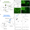
Our central hypothesis. (A) Proposed epileptic networks in patients with gelastic seizures associated with hypothalamic hamartoma (HH). Functional neuroimaging studies provided the evidence of excessive neural excitations in the lateral hypothalamus and mediodorsal thalamus during the ictal and interictal periods., Ablative disconnection between HH and the adjacent healthy brain resulted in long‐term control of gelastic seizures and improved cognitive function., (B) We hypothesized that local field potential (LFP) recording would clarify physiological neural activity supporting successful working memory formation. We also hypothesized that interictal epileptiform discharges (IEDs) propagating from the HH to critical brain structures during a critical period would result in transient impairment in working memory formation. (C) We employed channel rhodopsin‐2 photostimulation to the axon terminals from the lateral hypothalamus to the mediodorsal thalamus and prelimbic cortex in adult Long‐Evans rats. Photostimulation induced excessive neural excitation resembling IEDs in a time‐ and location‐specific manner. In the present study, we specifically determined whether the critical structures included the mediodorsal thalamus or prelimbic cortex, , , and whether IED occurrence during the encoding or maintenance period was critical. Figure 2E presents the explicit definitions of the pre‐encoding, encoding, and maintenance periods.
FIGURE 2
FIGURE 2
Outlines the experimental design. (A) A rat skull diagram displays the surgical plan for implanting a head fixation device, optrode, and chronic local field potential (LFP) recording electrodes, indicating anchor screw sites and a reference site for LFP recording. Br, bregma; L, lateral hypothalamic area; Lm, lambdoid suture; M, mediodorsal thalamus; and P, prelimbic cortex. (B) Illustration depicts a head‐fixed rat engaged in spout‐lever manipulation using a stereotaxic head fixation device for operant learning and LFP recordings during active and sham photostimulation. (C) The working memory task timeline involves a visual cue (green light‐emitting diode [LED] flashing) indicating the correct side (encoding period), followed by a randomly timed maintenance period and a “go” cue for lever‐pulling to obtain water rewards. (D) Photostimulation order: Rats undergo 20 discrimination task trials under sham and active photostimulation and 30 working memory task trials under both conditions. (E) Four photo‐stimulus conditions are defined based on period during tasks. (F) A daily schedule for assigning photostimulation patterns during the test days. The photostimulation patterns were fixed on the same day. (F) Daily schedule for assigning fixed photostimulation patterns during test days. Photostimulation effects are assessed by comparing success rates under active vs sham conditions. Stim., photostimulation.
FIGURE 3
FIGURE 3
Task performance and supporting neural activity. (A) Black dot indicates each rat's average success rate (%) in the task with a given maintenance period duration. The discrimination task was designed to have no maintenance period, whereas the working memory task was employed with a varying maintenance period (either 3, 5, 7, or 9 s). Under sham photostimulation, the success rate was lower in tasks with a longer maintenance period (mixed model‐based estimate [r] = −.66, p < .001). (B, C) The time–frequency maps time‐locked to the working memory encoding period offset show significant amplitude modulations (cluster‐based permutation tests, n = 1000, cluster size threshold α = .05) associated with successful trials (B) in the mediodorsal thalamus and (C) in the ipsilateral prelimbic cortex. Z‐scores below the significance threshold are masked. (D, E) Time–frequency maps demonstrate significant absolute beta coefficient values in the dorsomedial thalamus and ipsilateral prelimbic cortex. Beta coefficient values were computed based on a linear least absolute shrinkage and selection operator (LASSO)–support vector machine (LASSO‐SVM) model, which predicted successful or failed trials. The LASSO‐SVM models employed a fivefold cross‐validation approach. Beta coefficient values below the significance threshold are masked (nonparametric permutation testing, n = 1000, α = .05). Each cluster was numbered (see Figure S2).
FIGURE 4
FIGURE 4
The Channelrhodopsin‐2 (ChR2)‐expressing and the neural response to photo stimuli. (A–C) Scheme of virus injection. d3V, dorsal third ventricle; fs, fornix; Ic, internal capsule; IMD, Intermediodorsal thalamic nucleus; LHA, lateral hypothalamus area; LHb, lateral habenular nucleus; MD, thalamic mediodorsal nucleus (MDC, central part; MDL, lateral part; MDM, medial part); v3V, ventral third ventricle; ZI, zona incerta. (B) The filled‐blue circle indicates the position of the optrode tip. (D) The fluorescent micrograph in the virus injection area is shown in the illustrative schema (C). Arrows indicate ChR2‐Green fluorescent protein (GFP)‐expressing neural cell bodies in the LHA. The scale bar represents 80 μm. (E, F) Fluorescent micrographs in the photo‐stimulation area are shown in the illustrative schema (B). The ChR2‐expressing axon terminals in (E), MDL and (F) MDM; IMD. Insets in (E, F) are higher magnification of rectangular areas. Arrowheads indicate axonal varicosity in the area of mediodorsal thalamus. The scale bar represents 40 μm in (E, F). (G) Scheme of photostimulation at mediodorsal thalamus and local field potential (LFP) recording at the mediodorsal thalamus and ipsilateral prelimbic cortex (iPrL). (H–K) Grand average (n = 10 ChR2‐expressing rats) of the photo‐evoked responses (H, I) in the dorsomedial thalamus and (J, K) the iPrL. The baseline period for z‐transformation was −1.0 to −0.2 s for the photostimulus onset. Excitation photostimulus pattern: strength, 8 mW; Λ, 470 nm; pulse width, 500 μs, frequency, 80 Hz. (H) The blue bar indicates the photostimulus duration. (I) The blue arrowhead indicates the onset of a photo pulse, and the black arrowhead indicates the detected negative peak following that pulse. (J, K) Photostimulation at the dorsomedial thalamus elicited a thalamocortical evoked potential at the iPrL.
FIGURE 5
FIGURE 5
The task performance under the photostimulation. (A) An example of daily behavioral performance under the photostimulation during the encoding period. The success rate was defined as (100 × number of successful trials)/(number of successful trials + number of failure trials). The difference in the success rate (ΔSR) was defined as the difference between the success rate under active and sham photostimulation on a given day. (B) Photostimulation during the encoding period impaired the success rate. The degree of performance deterioration due to photostimulation during the encoding period was greater in the working memory task than in the discrimination task. The violin shape shows data distribution via kernel density estimation. The box represents the interquartile range (IQR), with the top and bottom indicating the first and third quartiles (Q1 and Q3). The line inside marks the median. Whiskers cover data within 1.5 times the IQR, and points beyond are outliers. Error bars indicate standard error (SE). * False discover rate (FDR)–corrected p‐value <.05; **< .01; ***< .001. (C) The degree of performance deterioration due to photostimulation during the encoding period was negatively correlated with the maintenance period duration in the working memory task. (D) A significant decrease in working memory task performance was observed with photostimulation, if active during the encoding period (i.e., encoding period alone: Red bar; encoding and maintenance periods: Purple bar). The statistical details are provided in Table S1. The violin shape shows data distribution via kernel density estimation. The box represents the IQR, with the top and bottom indicating the Q1 and Q3. The line inside marks the median. Whiskers cover data within 1.5 times the IQR, and points beyond are outliers. Error bars present SE. *FDR‐corrected p‐value < .05; *** < .001. The success rates for sessions with and without photostimulation are presented in Figure S6. (E) On a trial‐by‐trial analysis, negative predictors of trial success included photostimulation during either or both the encoding and maintenance periods and a longer maintenance period duration. The probability of success was marginally but significantly increased when task switch was required; the statistical details are provided in Table S2. Error bars indicate SE. *p‐value <.05. (F) The task performance under sham stimulation was compared between three periods (i.e., Days 1–2 vs Day 7 vs Day 12 in Figure 2F). There were no statistically significant differences in task performance among these periods.
FIGURE 6
FIGURE 6
Photostimulation‐induced alteration in working memory–related neural activity predicted failed trials. (A, B) The time–frequency map visualizes which components of neural activity contribute to failed trials. (A) Mediodorsal thalamus. (B) Ipsilateral prelimbic cortex. The degree of each cluster's contribution to predicition of failed trials are colored using t‐statistics (*p < .05). The values were calculated by mixed logistic regression analysis on the average local field potential (LFP) z‐value within each time–frequency cluster when photostimulation was applied during the encoding (i.e., latency from −1 to 0 s) and maintenance (latency from 0 to +2.5 s) periods, respectively. Statistical results are presented in detail (Table S3). Larger degrees of LFP amplitude augmentation (arrow, Cluster 3) and LFP amplitude attenuation (arrowhead, Cluster 8) under active photostimulation during the encoding period at the mediodorsal thalamus increased the probability of a given trial being failed.

References

    1. Kural MA, Duez L, Sejer Hansen V, Larsson PG, Rampp S, Schulz R, et al. Criteria for defining interictal epileptiform discharges in EEG: a clinical validation study. Neurology. 2020;94(20):e2139–e2147. - PMC - PubMed
    1. Holmes GL, Lenck‐Santini PP. Role of interictal epileptiform abnormalities in cognitive impairment. Epilepsy Behav. 2006;8(3):504–515. - PubMed
    1. Kleen JK, Scott RC, Holmes GL, Roberts DW, Rundle MM, Testorf M, et al. Hippocampal interictal epileptiform activity disrupts cognition in humans. Neurology. 2013;81(1):18–24. - PMC - PubMed
    1. Ung H, Cazares C, Nanivadekar A, Kini L, Wagenaar J, Becker D, et al. Interictal epileptiform activity outside the seizure onset zone impacts cognition. Brain. 2017;140(8):2157–2168. - PMC - PubMed
    1. Henin S, Shankar A, Borges H, Flinker A, Doyle W, Friedman D, et al. Spatiotemporal dynamics between interictal epileptiform discharges and ripples during associative memory processing. Brain. 2021;144(5):1590–1602. - PMC - PubMed