. 2025 Mar 11;20(3):e0310482.
doi: 10.1371/journal.pone.0310482. eCollection 2025.

Porphyromonas gingivalis outer membrane vesicles alter cortical neurons and Tau phosphorylation in the embryonic mouse brain

Affiliations

Porphyromonas gingivalis outer membrane vesicles alter cortical neurons and Tau phosphorylation in the embryonic mouse brain

Adrienne J Bradley et al. PLoS One. .

Abstract

Porphyromonas gingivalis (Pg) is an oral bacterial pathogen that has been associated with systemic inflammation and adverse pregnancy outcomes such as low birth weight and pre-term birth. Pg drives these sequelae through virulence factors decorating the outer membrane that are present on non-replicative outer membrane vesicles (OMV) that are suspected to be transmitted systemically. Given that Pg abundance can increase during pregnancy, it is not well known whether Pg-OMV can have deleterious effects on the brain of the developing fetus. We tested this possibility by treating pregnant C57/Bl6 mice with PBS (control) and OMV from ATCC 33277 by tail vein injection every other day from gestational age 3 to 17. At gestational age 18.5, we measured dam and pup weights and collected pup brains to quantify changes in inflammation, cortical neuron density, and Tau phosphorylated at Thr231. Dam and pup weights were not altered by Pg-OMV exposure, but pup brain weight was significantly decreased in the Pg-OMV treatment group. We found a significant increase of Iba-1, indicative of microglia activation, although the overall levels of IL-1β, IL-6, TNFα, IL-4, IL-10, and TGFβ mRNA transcripts were not different between the treatment groups. Differences in IL-1β, IL-6, and TNFα concentrations by ELISA showed IL-6 was significantly lower in Pg-OMV brains. Cortical neuron density was modified by treatment with Pg-OMV as immunofluorescence showed significant decreases in Cux1 and SatB2. Overall p-Tau Thr231 was increased in the brains of pups whose mothers were exposed to Pg-OMV. Together these results demonstrate that Pg-OMV can significantly modify the embryonic brain and suggests that Pg may impact offspring development via multiple mechanisms.


Conflict of interest statement

The authors have declared that no competing interests exist.

Figures

Fig 1
Fig 1. Dam and pup body and brain weights.
(A) and (B) Weight of dams and pups were not significantly different from PBS controls. (C) Pup brain weight decreased significantly with Pg exposure (Mann-Whitney U p =  0.023). (D) Relative expression of Hif1a as indicator of hypoxia in the pup brain. Relative expression calculated by the 2^-(ΔΔCt) method with GAPDH as the reference gene. Data are shown as median with 95% CI.
Fig 2
Fig 2. Increased Iba-1 in mouse pups exposed to Pg-OMV during pregnancy.
(A) Western blot of Iba-1 and β-actin in GA 18.5 pups from PBS and Pg-OMV exposed dams. Full blot can be found in the Supporting information. (B) Comparison of β-actin normalized signal intensity of Iba-1 showed significant increase in Iba-1 in the Pg-OMV group compared to controls (Mann-Whitney U p =  0.0003). Data are shown as median with 95% CI.
Fig 3
Fig 3. ELISA and RT-qPCR evaluation of cytokines in the embryonic mouse brain.
(A, D, G): ELISA of IL-1β, IL-6, and TNFα in pg ml-1. Cytokines were extracted from frozen whole brains and quantified. IL-1β was significantly greater in the PBS female pups compared to the female pups from the Pg-OMV group (Fig 3A, Mann-Whitney U test p =  0.04). IL-6 was significantly greater in the PBS group and in the female pups compared to the OMV treatment group (Fig 3D, Student’s T-test p =  0.002 and 0.0095, respectively.) (B, E, H): RT-qPCR of proinflammatory cytokines IL-1β, IL-6, and TNFα. (C, F, I): RT-qPCR of anti-inflammatory cytokines IL-4, IL-10, and TGFβ. Total RNA was extracted from frozen whole brains and reverse transcribed to cDNA. Relative expression calculated by the 2^-(ΔΔCt) method with GAPDH as the reference gene. Group differences for the ELISAs on the males was not determined due to low sample size (n =  2). Only significant differences are shown. Data are presented with the median and 95% CI. Symbols: Circles =  PBS, triangles =  Pg; orange =  females; blue =  males.
Fig 4
Fig 4. RT-qPCR evaluation of MYD88, NLRP3, and NFκβ in GA 18.5 pup brains.
(A) MyD88 and (B) NLRP3 were not significantly different in pup brains from the control or treatment group. (C) NFkβ was significantly reduced in the pup brains (Student’s T-test p =  0.004). Total RNA was extracted from frozen whole brains and reverse transcribed to cDNA. Relative expression calculated by the 2^-(ΔΔCt) method with GAPDH as the reference gene. Means across fore, mid, and hind brain sections are shown for each sample. Data are presented with the median value and 95% CI.
Fig 5
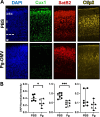
Fig 5. Modified intensity and location of cortical layer marker proteins following Pg-OMV exposure in utero.
(A) Representative immunofluorescent images from sagittal sections of PFA fixed brains at GA 18.5 pup brains showing location of cortical layer markers Cux1, SatB2, and Ctip2. Approximate position of cortical layers 1–6 with the intraventricular zone (IZ) are shown in the PBS-DAPI image. (B) Quantification and comparison of cortical layer proteins stained with the marker antibodies. Intensities were averaged across three serial section per sample for statistical analysis. Cux1 (Student’s T-test p =  0.03) and SatB2 (students T-test p =  0.0001) were significantly reduced in the Pg-OMV group. Data are presented with the median and 95% CI.
Fig 6
Fig 6. Differences in total Tau and p-Tau Thr231 following maternal Pg-OMV exposure.
(A-B) Representative images of WB showing total Tau and p-Tau Thr231 Tau from E18.5 pup brains. (C-D) Quantification of western blots showing decrease in total Tau and increase in p-Tau Thr231 (Student’s T-Test p =  0.0013). (E) Ratio of p-Tau to Total Tau was significantly increased in the Pg pups (Mann-Whitney U Test p =  0.0025). Data show mean + /- SEM.

References

    1. Carrillo-de-Albornoz A, Figuero E, Herrera D, Bascones-Martínez A. Gingival changes during pregnancy: II. influence of hormonal variations on the subgingival biofilm. J Clin Periodontol. 2010;37(3):230–40. doi: 10.1111/j.1600-051X.2009.01514.x - DOI - PubMed
    1. Fujiwara N, Tsuruda K, Iwamoto Y, Kato F, Odaki T, Yamane N, et al.. Significant increase of oral bacteria in the early pregnancy period in Japanese women. J Investig Clin Dent. 2017;8(1):e2189. doi: 10.1111/jicd.12189 - DOI - PubMed
    1. Lin W, Jiang W, Hu X, Gao L, Ai D, Pan H, et al.. Ecological shifts of supragingival microbiota in association with pregnancy. Front Cell Infect Microbiol. 2018;8:24. doi: 10.3389/fcimb.2018.00024 - DOI - PMC - PubMed
    1. Paropkari AD, Leblebicioglu B, Christian LM, Kumar PS. Smoking, pregnancy and the subgingival microbiome. Scientific Reports. 2016;6(1):30388. - PMC - PubMed
    1. Jang H, Patoine A, Wu T, Castillo D, Xiao J. Oral microflora and pregnancy: a systematic review and meta-analysis. Scientific Reports. 2021;11(1):16870. - PMC - PubMed

LinkOut - more resources