. 2025 Mar 18;122(11):e2423931122.
doi: 10.1073/pnas.2423931122. Epub 2025 Mar 11.

Epitope-directed selection of GPCR nanobody ligands with evolvable function

Affiliations

Epitope-directed selection of GPCR nanobody ligands with evolvable function

Meredith A Skiba et al. Proc Natl Acad Sci U S A. .

Abstract

Antibodies have the potential to target G protein-coupled receptors (GPCRs) with high receptor, cellular, and tissue selectivity; however, few antibody ligands for GPCRs exist. Here, we describe a generalizable selection method to enrich for GPCR ligands from a synthetic camelid antibody fragment (nanobody) library. Our strategy yielded multiple nanobody ligands for the angiotensin II type I receptor (AT1R), a prototypical GPCR and important drug target. We found that nanobodies readily act as allosteric modulators, encoding selectivity for both the receptor and chemical features of GPCR ligands. We then used structure-guided design to convert two nanobodies from allosteric ligands to competitive AT1R inhibitors through simple mutations. This work demonstrates that nanobodies can encode multiple pharmacological behaviors and have great potential as evolvable scaffolds for the development of next-generation GPCR therapeutics.

Keywords: GPCR; allostery; angiotensin; cryo-EM; nanobody.


Conflict of interest statement

Competing interests statement:A.C.K. is a cofounder and consultant for biotechnology companies Tectonic Therapeutic and Seismic Therapeutic, and also for the Institute for Protein Innovation, a nonprofit research institute. A.C.K. holds stock in Tectonic Therapeutic and Seismic Therapeutic.

Figures

Fig. 1.
Fig. 1.
Identification of nanobody ligands for AT1R. (A) Flowchart of the selection process. Extracellular-binding nanobodies were enriched from a synthetic nanobody library through two rounds of MACS. In the third round of selection, nanobodies that preferentially bound the extracellular surface of AT1R were sorted via FACS. (B) Cartoon of the FACS selection step. Only the extracellular surface of AT1R is accessible for yeast-displayed nanobody binding to the FLAG-AT1R-AT110d4 fusion protein. Yeast-displayed nanobodies binding to the intracellular surface or nanobody framework bind to the AT118-H-AT1R-Protein C fusion protein. Both fusion proteins were labeled with different fluorophore dye-conjugated antibodies for fluorescence-activated cell sorting. The yeast population that preferentially bound to FLAG-AT1R-AT110d4 was collected. (C) Binding of single yeast-displayed nanobody clones to 100 nM unliganded, AngII, and antagonist-bound AT1R in detergent. All nanobodies bound to unliganded AT1R and competed for binding with AngII. Variable binding was observed in the presence of AT1R antagonists. Data are presented as mean ± SEM from three experiments. (D) CDR sequences of identified nanobodies. Bolded residues were diversified positions in the nanobody library (20). (E) Binding of 250 nM purified nanobodies to unliganded, AngII, and antagonist-bound AT1R in intact cell membranes. Nine of 11 nanobodies bound to the extracellular surface of AT1R. Many nanobodies display preferential binding for the antagonist-bound receptor. Data are presented as mean ± SEM from three experiments.
Fig. 2.
Fig. 2.
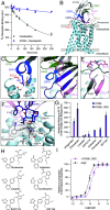
Characterization and engineering of AT209. (A) AT209 effects on the dissociation of [3H]-olmesartan from AT1R in cell membranes. Data are presented as mean ± SEM from three experiments. AT209 slows the off-rate of olmesartan, indicating that AT209 allosterically potentiates olmesartan binding to AT1R. (B) Cryo-EM structure of AT209 in complex with olmesartan-bound AT1R. AT209’s CDRs interact with AT1R’s ECLs. Image is colored by component AT209 (gray), CDR1 (dark blue), CDR2 (light green), CDR3 (purple), AT1R (light blue), ECL1 (yellow), ECL2 (light pink), and ECL3 (red). (C) AT209 CDR1 (blue) interacts with acidic residues on TMs 6 and 7. Dashed lines represent hydrogen bonds. (D) AT209 CDR3 (purple) forms a continuous β-sheet with AT1R’s ECL2 (light pink). This interaction is stabilized by CDR2 (light green). (E) In active-state AT1R structures, AngII (gray) forms a continuous β-sheet with AT1R’s ECL2 (light pink), which is supported by the receptors N terminus (turquoise). (F) AT209 CDR3 (purple) sits within AT1R’s olmesartan-bound (gray sticks) orthosteric pocket. L103CDR3 sits within a hydrophobic pocket. (G) Binding of 250 nM AT209 variants to antagonist-bound AT1R in intact cells. (H) Chemical structures of AT1R antagonists. (I) AT209 L103D suppress AngII-mediated Gαq signaling, as measured through the NFAT transcriptional reporter. Data are presented as mean ± SEM from three experiments.
Fig. 3.
Fig. 3.
Characterization of AT206. (A) AT206 potentiates the inhibitory activity of losartan on AngII-mediated Gαq signaling, as measured through the production of IP3. Data are presented as mean ± SEM from three experiments. (B) Cryo-EM structure of AT206 in complex with losartan-bound AT1R. CDR3 enters AT206’s orthosteric pocket, while regions of CDRs 1 and 2 are disordered (dashed lines). Image is colored by component AT206 (gray), CDR1 (dark blue), CDR2 (light green), CDR3 (orange), AT1R (light blue), ECL1 (yellow), ECL2 (light pink), and ECL3 (red). (C) AT206 CDR3 (orange) forms a continuous β-strand with ECL2 (light pink). This interaction is supported by CDR2 (light green). Black dashed lines indicate hydrogen bonds. (D) CDR3 (orange) interacts with AT1R’s ECL1 (yellow) and TMs 5, 6, and 7. (E) The backbone of V105CDR3 forms a hydrogen bond network (black dashed lines) with losartan and residues within AT1R’s orthosteric pocket. (F) Comparison of AT206 CDR3 in Cryo-EM structures of losartan-bound (orange) and unliganded (purple) AT1R. CDRs 1 and 2 are colored as in (B). (G) In unliganded AT1R, AT206 CDR3 (purple) occupies losartan’s binding site.
Fig. 4.
Fig. 4.
Engineering of AT206. (A). Overlay of AT206, AT209, and AT118-bound AT1R. All three CDRs are oriented similarly. Image is colored as follows: CDR1 (AT206, orange; AT209, purple; AT118, dark blue), CDR2 (light green), CDR3 (magenta), AT1R (light blue), ECL1 (yellow), ECL2 (light pink), and ECL3 (red). Unlike the allosteric parent nanobody AT206, the AT118-AT206 chimera suppresses AngII-mediated (B) Gαq signaling, as measured through the NFAT transcriptional reporter and (C) β-arrestin 2 recruitment, as measured through the TANGO assay. Signaling assay data are presented as mean ± SEM from three experiments.

References

    1. Santos R., et al. , A comprehensive map of molecular drug targets. Nat. Rev. Drug Discov. 16, 19–34 (2017). - PMC - PubMed
    1. Hutchings C. J., Koglin M., Olson W. C., Marshall F. H., Opportunities for therapeutic antibodies directed at G-protein-coupled receptors. Nat. Rev. Drug Discov. 16, 787–810 (2017). - PubMed
    1. Jähnichen S., et al. , CXCR4 nanobodies (VHH-based single variable domains) potently inhibit chemotaxis and HIV-1 replication and mobilize stem cells. Proc. Natl. Acad. Sci. U.S.A. 107, 20565–20570 (2010). - PMC - PubMed
    1. Maussang D., et al. , Llama-derived single variable domains (nanobodies) directed against chemokine receptor CXCR7 reduce head and neck cancer cell growth in vivo. J. Biol. Chem. 288, 29562–29572 (2013). - PMC - PubMed
    1. Scholler P., et al. , Allosteric nanobodies uncover a role of hippocampal mGlu2 receptor homodimers in contextual fear consolidation. Nat. Commun. 8, 1967 (2017). - PMC - PubMed

MeSH terms

LinkOut - more resources