. 2025 Mar 11;18(877):eadr0128.
doi: 10.1126/scisignal.adr0128. Epub 2025 Mar 11.

NEMO is essential for directing the kinases IKKα and ATM to the sites of DNA damage

Affiliations

NEMO is essential for directing the kinases IKKα and ATM to the sites of DNA damage

Josune Alonso-Marañón et al. Sci Signal. .

Abstract

The DNA damage repair kinase ATM is phosphorylated by the NF-κB pathway kinase IKKα, resulting in enhanced DNA damage repair through the nonhomologous end-joining pathway. Thus, inhibition of IKKα enhances the efficacy of cancer therapy based on inducing DNA damage. Here, we found a role for the IKK regulatory subunit NEMO in DNA damage repair mediated by ATM and IKKα. Exposure to damaging agents induced the interaction of NEMO with a preformed ATM-IKKα complex, which was required to target active ATM and IKKα to chromatin for efficient DNA damage repair but not for activating ATM. Recognition of damaged DNA by the IKKα-NEMO-ATM complex was facilitated by the interaction between NEMO and histones and depended on the ADP ribosylation of histones by the enzyme PARP1. NEMO-deficient cells showed increased activity of the kinase ATR, and inhibition of ATR potentiated the effect of chemotherapy in cells lacking NEMO or IKKα. Bioinformatic analysis of colorectal cancer datasets demonstrated that the expression of genes encoding IKKα, NEMO, and ATM correlated with poor patient prognosis, suggesting that the mechanism linking these three elements may be clinically relevant.


Conflict of interest statement

Competing interests: The authors declare that they have no competing financial interests related to this work.

Figures

Fig. 1.
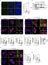
Fig. 1.. Dynamic association of IKKα ad NEMO to DDR proteins in response to UV light treatment.
(A) Representation of the results obtained in the mass spectrometry analysis of streptavidin precipitates from HEK293T cells expressing the fusion protein BirA-IKKα, pre-treated overnight with biotin (50 μM), and either untreated or exposed for 15 or 60 min to 130 mJ/cm2 UV light. Analysis shown represents the mean of the results obtained from 3 biological replicates. (B) Network of selected proteins identified as putative IKKα interactors in cells described in (A). (C) Soluble extracts from HEK293T BirA-IKKα cells, pre-treated overnight with biotin (50 μM) and either untreated (right) or exposed for 30 min to 130 mJ/cm2 UV light (left) were fractionated in Superdex200 columns. Two drops per fraction were collected and precipitated with streptavidin-coated beads, and the abundance of ATM, IKKα, and NEMO were assessed by WB. Blots are a representative of 3 biological replicates. The fraction in which the 200 kDa molecular marker was eluted (fraction 6) is marked. (D and E) WB analysis of streptavidin precipitates from soluble (D) or chromatin (E) extracts of HEK293T cells transfected with BirA-IKKα plasmid, pretreated overnight with biotin (50 μM) and exposed to UV light (130 mJ/cm2) for 15 or 60 min. Blots are from a representative of 3 biological replicates. (F and G) PLA using antibodies against phosphorylated (p-) IKKα and IKKβ and γH2A.X (F) or NEMO and γH2A.X (G) was assessed in Caco2 cells treated with 50 μg/mL 5-FU and 20 μg/mL irinotecan (5-FU+Iri.) for 16 hours. As negative controls, the PLA reaction following incubation with single antibodies was assessed and is shown (“-only”). Images are representative, and quantification of the number of PLA dots per cell in the indicated conditions is shown as the means and standard error of mean (SEM) from n >300 cells from > 3independent experiments. ****p <0.0001 by unpaired two-tailed t-tests. Scale bars: 25 μm, 25 μm and 10 μm from left to right. (H) WB analysis of streptavidin precipitates of total extracts from HCT116 cells expressing BirA-H2A.X protein, pre-treated overnight with biotin (50 μM), then either untreated or UV light-exposed (130 mJ/cm2 for 30 min). Blots are from a representative of 3 biological replicates.
Fig. 2.
Fig. 2.. Defective nuclear distribution of IKKα and ATM in NEMO-deficient cells.
(A) WB analysis of chromatin extracts from Caco2 WT and NEMO KO cells exposed to UV light (130 mJ/cm2) and collected at the indicated time points (1 representative of 3 biological replicates). (B) Representative images of ATM staining in NEMO WT and NEMO KO Caco2 cells untreated and exposed to UV light (130 mJ/cm2) for 30 minutes (1 representative of 3 biological replicates). Scale bars: 25 μm, 25 μm and 10 μm, from left to right. (C) Quantification of the percentage of positive cells displaying nuclear ATM described in (B), graphs representing the means and SEM from n=6 biological replicates. ****p <0.0001, ***p <0.001, n.s. not significant, by one-way ANOVA. (D) Representative images of ATM staining in NEMO WT and NEMO KO Caco2 cells untreated or pretreated with Leptomycin B for 16 hours (5μg/μL) and then left untreated or exposed to UV light for 30 minutes (130 mJ/cm2). Images are from 1 of 3 biological replicates. Scale bars: 25 μm. (E) Quantification of the percentage of cells showing the indicated subcellular distribution of ATM described in (D), graphs representing the means from n=3 20X magnification fields counted from 3 biological replicates. (F) WB analysis of streptavidin precipitates from chromatin extracts of WT or NEMO KO HEK293T cells carrying the BirA-IKKα fusion protein, pretreated with Biotin (50μM, O/N) and treated with UV light (130 mJ/cm2) as indicated. Blots are from 1 representative experiment from 3 biological replicates. Input represents 5% of the lysate. (G) Comet assay from Caco2 WT and NEMO KO treated with the combination of 5-FU+Irinotecan (5μg/mL+2μg/mL) for 24 and 72 hours. Graphs represent the means and SEM from n >400 cells examined over 3 independent experiments. ****p <0.0001, ***p <0.001 by one-way ANOVA. (H) Dose-response assay using the chemotherapeutic combination of 5-FU+Irinotecan in Caco2 WT and NEMO KO cells at indicated concentrations for 72 hours, graphs representing the means and SEM from n=3 biological replicates. P-values were derived from a two-sided paired t-test, comparing treated with untreated condition and treated conditions with each other.
Fig. 3.
Fig. 3.. NEMO regulates chromatin association of IKKα and active ATM to sites of DNA damage.
(A) PLA with antibodies against phosphorylated (p-)IKKα and IKKβ and γH2A.X of WT and NEMO KO Caco2 cells treated with 5-FU+Irinotecan (50μg/mL+20μg/mL) for 16 hours and quantification of the number of PLA dots per cell in the indicated conditions, graphs representing means and SEM from n = more than 400 cells examined over three independent experiments. ****p <0.0001 by one-way ANOVA. Scale bars, left to right: 25, 25, and 10 μm. (B) WB analysis of streptavidin precipitates of total extracts from WT and NEMO KO HCT116 cells expressing BirA-H2A.X, pretreated with Biotin (50μM, O/N), and left untreated or treated with UV light (130 mJ/cm2) for 1 hour. Blots are from 1 representative of 3 biological replicates. (C to F) Double IF analysis of p-ATM and γH2A.X in Caco2 WT and NEMO KO cells exposed to UV light (130 mJ/cm2, 30min) (1 representative of 3 biological replicates is shown) (C) and quantification of the percentage of cells treated with UV light positive for nuclear p-ATM (D) and γH2A.X (E), and percentage of cells with co-localizing p-ATM and γH2A.X dots (F). Graphs represent means and SEM from a minimum of n=7 20X fields from 3 biological replicates. ****p <0.0001, ***p <0.001, **p <0.01, n.s. not significant, by one-way ANOVA. Scale bars, left to right: 25, 25, 10, and 10 μm. (G to J) Double IF analysis of p-ATM and γH2A.X in Caco2 WT and NEMO KO cells treated with 5-FU+Irinotecan (50 μg/mL+20 μg/mL) for 16 hours (G) and quantification of the percentage of cells displaying nuclear p-ATM (H), double-positive for p-ATM and γH2A.X (I), and number of co-localizing dots per nucleus (J). Graphs represent means and SEM from a minimum of n=3 20X fields from 3 biological replicates. ****p <0.0001, **p <0.01 by one-way ANOVA. Scale bars in (G), left to right: 25, 25, 10, and 10 μm. (K to M) Double IF analysis of p-53BP1 and γH2A.X in Caco2 WT and NEMO KO cells treated with 5-FU+Irinotecan for 16 hours (K) and quantification of cells carrying nuclear p-53BP1 (L) and colocalizing p-53BP1 and γH2A.X dots (M). Graphs represent means and SEM from n=2, 4, and 5 20X fields from 3 biological replicates. ***p <0.001, **p <0.01, n.s. not significant by one-way ANOVA. Scale bars, left to right: 25, 25, 10, and 10 μm.
Fig. 4.
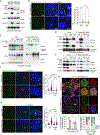
Fig. 4.. Recruitment of IKKα and phosphorylated ATM to the damaged DNA is NEMO and PARP1 dependent.
(A) WB analysis of chromatin extracts from HCT116 NEMO KO cells expressing MT-NEMO WT or MT-NEMO lacking the C-terminal zinc finger domain (ΔZF) treated as indicated with UV light (130 mJ/cm2). Blots are from 1 representative of 3 biological replicates. (B) PLA with antibodies against p-IKKα and IKKβ and γH2A.X of NEMO KO HCT116 cells expressing MT-NEMO WT or MT-NEMO ΔZF and treated with 5-FU+Irinotecan (50μg/mL+20μg/mL) for 16 hours. Quantification of the number of PLA dots per cell, graphs representing means and SEM from n >400 cells examined over 3 independent experiments. ****p <0.0001, **p <0.01 by one-way ANOVA. Scale bars, left to right: 25, 25, and 10 μm. (C and D) WB analysis of streptavidin precipitates of chromatin extracts from WT and NEMO KO (C) or PARP1 KO (D) HEK293T cells expressing BirA-IKKα plasmid, pretreated with biotin (50 μM overnight), and left untreated or treated with UV light (130 mJ/cm2) as indicated. Blots are from 1 representative of 3 biological replicates. (E) WB analysis of streptavidin precipitates of total extracts from WT and PARP1 KO HCT116 cells expressing BirA-H2A.X, pretreated with biotin (50 μM overnight), and left untreated or treated with UV light (130 mJ/cm2) for 1 hour. Blots are from 1 representative of 3 biological replicates. (F and G) PLA with antibodies against p-IKKα and IKKβ and γH2A.X (F) or NEMO and γH2A.X (G) in WT and PARP1 KO Caco2 cells either untreated or treated with 5-FU (50 μg/mL) and Irinotecan (20 μg/mL) for 16 hours. Quantification of the number of PLA dots per cell is shown, from n >300 cells examined over 3 independent experiments. ****p <0.0001 by one-way ANOVA. Scale bars, left to right: 25, 25, and 10 μm. (H to J) IF analysis of phosphorylated ATM (p-ATM) and γH2A.X in the cells described and as treated in (F and G), with representative images shown (H), the number of cells double-positive for p-ATM and γH2A.X quantified (I), and the percent of cells with the indicated number of colocalizing dots assessed (J). Graphs show means and SEM from a minimum of n=3 20X fields from 3 biological replicates. ***p <0.001 and **p <0.01 by one-way ANOVA. Scale bars in (H), left to right: 25, 10, and 10 μm.
Fig. 5.
Fig. 5.. High levels of IKBKG (NEMO) are predictive of poor prognosis in CRC patients.
(A and B) Kaplan–Meier representation of DFS over time for patients of all stages or stages II+III according to IKBKG levels (high or low relative to best cutoff) in the Marisa (A) and TCGA (B) colorectal cancer databases (number of patients indicated in the figure). For statistical analysis of the Kaplan-Meier estimates we used Cox proportional hazards models and log-rank two-sided test. HR, hazard ratio.; p, p-value. (C and D) Forest plot for the multivariate Cox proportional hazard regression model showing hazard ratio estimates and 95% confidence intervals from the IKBKG gene (codifying for NEMO) in Marisa (C) and TCGA (D) databases. Other parameters included in the analysis are indicated. NA: not ascribed. The number of patients (n) is indicated in the figure. (E) Kaplan–Meier representation of DFS over time for patients of stages II+III treated with neoadjuvant chemotherapy. The number of patients (n) is indicated in the figure. For statistical analysis of the Kaplan-Meier estimates we used Cox proportional hazards models and log-rank two-sided test. HR, hazard ratio.; p, p-value. (F and G) Enrichment pathway analysis of genes differentially expressed (down- or up-regulated) in the group of IKBKG-low tumors in ATM high (F) or CHUK high (G) patient subgroups from Marisa dataset. (H) Violin plots depicting ATR expression levels of tumors in the Marisa dataset classified according to IKBKG levels. For statistical analysis we used Wilcoxon rank-sum test. p, p-value. (I) WB analysis of phosphorylated (p-) and total ATR protein in WT and NEMO KO Caco2 cells treated with UV light (130 mJ/cm2) (top panel) and in HCT116 cells treated with 5-FU+Irinotecan (50 μg/mL + 20 μg/mL) (bottom panel) as indicated. Blots are from 1 representative of 3 biological replicates. (J to L) Dose-response assay of WT and NEMO KO Caco2 (J), HCT116 (K) and PDO5 cells (L) treated with ATR inhibitor as indicated for 72 hours. Graphs represent the means and SEM from n=3 biological replicates. Statistical analysis was performed by two-sided paired t-test, comparing treated with untreated condition and treated conditions with each other. (M to O) Dose-response assay of WT and NEMO KO Caco2 (M), HCT116 (N) and PDO5 cells (O) treated with 5-FU+Irinotecan as indicated in combination with ATRi (VE-821, 1μM, 0.5μM and 1.67μM respectively. Graphs represent the means and SEM from n=3 biological replicates. Statistical analysis was performed by two-sided paired t-test, comparing treated with untreated condition and treated conditions with each other.
Fig. 6.
Fig. 6.. Model for NEMO function at the chromatin after exposure of cells to damaging agents.
ATM and IKKα form a complex under basal conditions to allow rapid phosphorylation of ATM by IKKα in response to damage downstream of BRAF and p38α. Upon damage, NEMO binds to the activated ATM-IKK complex to promote nuclear translocation and recruitment of active ATM and IKK (most likely IKKα) to damaged DNA sites, probably through interaction with the DNA damage sensor PARP1. Recruitment of this complex to chromatin enables effective DNA repair and cell survival. In addition, PARP1 activity facilitates the recruitment of PIASy to the NEMO-ATM complex, resulting in NEMO SUMOylation and NF-kB signaling. Whether NEMO SUMOylation is required for IKKα-dependent DNA repair remains to be investigated. In the absence of NEMO, the activated ATM-IKK complex cannot associate with PARP1; therefore, it fails to recognize damaged DNA sites, resulting in defective DNA repair and NF-kB signaling leading to increased cell death. Defective NEMO activity is partially compensated by activation of alternative DNA repair and survival pathways. IKKα KO or its inhibition by the BRAF inhibitor AZ628 precludes activation of the ATM-IKK complex, thereby compromising its function in the DDR pathway.

Similar articles

Cited by

References

    1. Touil Y, Igoudjil W, Corvaisier M, Dessein A-F, Vandomme J, Monté D, Stechly L, Skrypek N, Langlois C, Grard G, Millet G, Leteurtre E, Dumont P, Truant S, Pruvot F-R, Hebbar M, Fan F, Ellis LM, Formstecher P, Van Seuningen I, Gespach C, Polakowska R, Huet G, Colon Cancer Cells Escape 5FU Chemotherapy-Induced Cell Death by Entering Stemness and Quiescence Associated with the c-Yes/YAP Axis. Clin. Cancer Res. 20, 837–846 (2014). - PMC - PubMed
    1. Dhimolea E, de Matos Simoes R, Kansara D, Al’Khafaji A, Bouyssou J, Weng X, Sharma S, Raja J, Awate P, Shirasaki R, Tang H, Glassner BJ, Liu Z, Gao D, Bryan J, Bender S, Roth J, Scheffer M, Jeselsohn R, Gray NS, Georgakoudi I, Vazquez F, Tsherniak A, Chen Y, Welm A, Duy C, Melnick A, Bartholdy B, Brown M, Culhane AC, Mitsiades CS, An Embryonic Diapause-like Adaptation with Suppressed Myc Activity Enables Tumor Treatment Persistence. Cancer Cell 39, 240–256.e11 (2021). - PMC - PubMed
    1. Rehman SK, Haynes J, Collignon E, Brown KR, Wang Y, Nixon AML, Bruce JP, Wintersinger JA, Singh Mer A, Lo EBL, Leung C, Lima-Fernandes E, Pedley NM, Soares F, McGibbon S, He HH, Pollet A, Pugh TJ, Haibe-Kains B, Morris Q, Ramalho-Santos M, Goyal S, Moffat J, O’Brien CA, Colorectal Cancer Cells Enter a Diapause-like DTP State to Survive Chemotherapy. Cell, doi: 10.1016/j.cell.2020.11.018 (2021). - DOI - PMC - PubMed
    1. Solé L, Lobo-Jarne T, Villanueva A, Vert A, Guillén Y, Sangrador I, Barbachano A, Lop J, Guix M, Salido M, Bellosillo B, García-Romero R, Garrido M, González J, Martínez-Iniesta M, Lopez-Arribillaga E, Salazar R, Montagut C, Torres F, Iglesias M, Celià-Terrassa T, Muñoz A, Bigas A, Espinosa L, Chemotherapy induces a YAP1-dependent fetal conversion to human Colorectal Cancer cells that is predictive of poor patient outcome. bioRxiv (2021).
    1. Cánovas B, Igea A, Sartori AA, Gomis RR, Paull TT, Isoda M, Pérez-Montoyo H, Serra V, González-Suárez E, Stracker TH, Nebreda AR, Targeting p38α Increases DNA Damage, Chromosome Instability, and the Anti-tumoral Response to Taxanes in Breast Cancer Cells. Cancer Cell, doi: 10.1016/j.ccell.2018.04.010 (2018). - DOI - PubMed

MeSH terms

LinkOut - more resources