. 2025 Mar 11;16(1):2436.
doi: 10.1038/s41467-025-57768-9.

The structural basis for the human procollagen lysine hydroxylation and dual-glycosylation

Affiliations

The structural basis for the human procollagen lysine hydroxylation and dual-glycosylation

Junjiang Peng et al. Nat Commun. .

Abstract

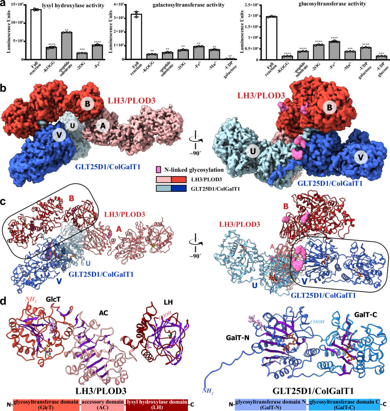
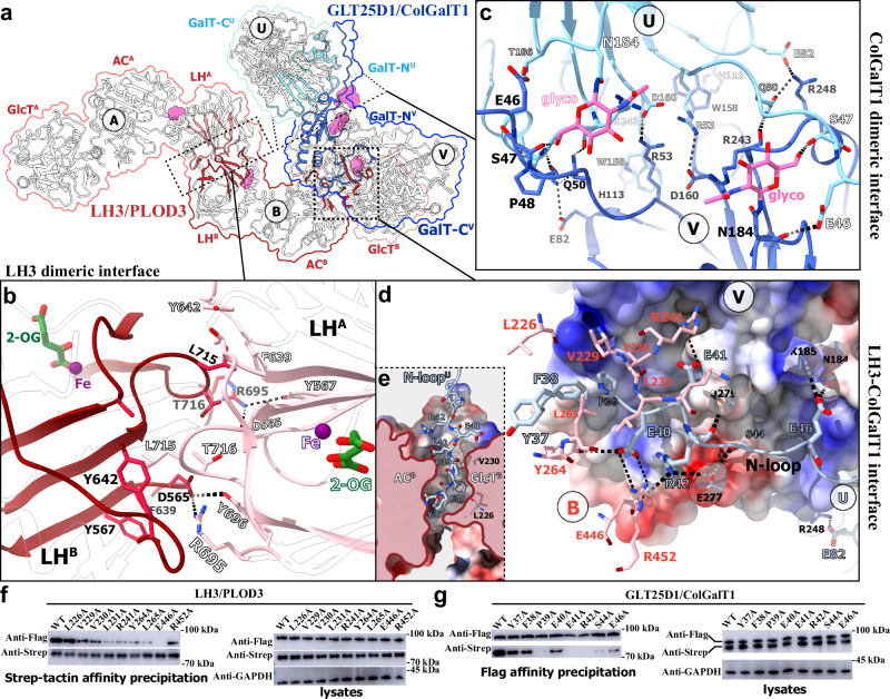
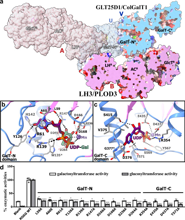
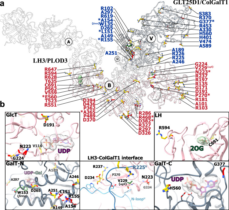
The proper assembly and maturation of collagens necessitate the orchestrated hydroxylation and glycosylation of multiple lysyl residues in procollagen chains. Dysfunctions in this multistep modification process can lead to severe collagen-associated diseases. To elucidate the coordination of lysyl processing activities, we determine the cryo-EM structures of the enzyme complex formed by LH3/PLOD3 and GLT25D1/ColGalT1, designated as the KOGG complex. Our structural analysis reveals a tetrameric complex comprising dimeric LH3/PLOD3s and GLT25D1/ColGalT1s, assembled with interactions involving the N-terminal loop of GLT25D1/ColGalT1 bridging another GLT25D1/ColGalT1 and LH3/PLOD3. We further elucidate the spatial configuration of the hydroxylase, galactosyltransferase, and glucosyltransferase sites within the KOGG complex, along with the key residues involved in substrate binding at these enzymatic sites. Intriguingly, we identify a high-order oligomeric pattern characterized by the formation of a fiber-like KOGG polymer assembled through the repetitive incorporation of KOGG tetramers as the biological unit.


Conflict of interest statement

Competing interests: The authors declare no competing interests.

Figures

Fig. 1
Fig. 1. The overall architecture of the human KOGG complex.
a Multi-functions of Purified LH3-ColGalT1 Complex. Left: 2-OG-dependent oxygenase activities of the purified KOGG complex. Oxygenase activities were minimal upon the removal of 2-OG from the reaction mixtures and substantially reduced in the absence of the KOGG complex sample, Fe2+, or substrate peptide. Middle: Galactosyltransferase activities of the purified KOGG complex. The galactosyltransferase activities were minimal upon the removal of UDP-galactose from the reaction mixtures and substantially reduced in the absence of the KOGG complex sample, Mn2+, or substrate peptide, as well as the necessary reaction components of 2-OG-dependent oxygenase activities, i.e., 2-OG and Fe2+. Right: Glucosyltransferase activities of the purified KOGG complex. The glucosyltransferase activities were minimal upon the removal of UDP-glucose from the reaction mixtures and also substantially reduced in the absence of the KOGG complex sample, Mn2+, or substrate peptide, as well as the necessary reaction components of 2-OG-dependent oxygenase activities, i.e. 2-OG and Fe2+, and the necessary reaction components of galactosyltransferase activities, UDP-galactose. (n = 3 biologically independent samples). Data are presented as mean values +/− SD. Source data are provided as a Source Data file. Data analysis was performed using one-way ANOVA with Dunnett’s multiple comparisons test. **, P-value < 0.01; ***, P-value < 0.001; ****, P-value < 0.0001. b Cryo-EM map of the heterotetrametic complex of human LH3 and ColGalT1. The cryo-EM density map of the human KOGG/UDP-Gal complex was viewed from two angles. The cryo-EM density map is colored in light red/red for LH3A/B, cyan/blue for ColGalT1U/V and magenta for glycosyl modifications. c Molecular model of the KOGG complex. KOGG/UDP-Gal complex was depicted as a cartoon model, colored by protomers according to the corresponding cryo-EM maps shown above. d Structural analysis on LH3 (left) and ColGalT1 (right). The structure of LH3B and ColGalT1V, indicated by the frames in the complex model above, are depicted as a cartoon model, colored by domains to the schemes shown below.
Fig. 2
Fig. 2. Molecular architecture of KOGG quaternary complex.
a Overview of the intermolecular contacts within the KOGG complex. The protomers are depicted as a cartoon model, with secondary structures involved in complex formation highlighted in pink/red for LH3 and in blue/cyan for ColGalT1. Interfaces mediating complex assembly indicated by dotted frames. The N-linked glycosylations were depicted as surface models in magenta. b Enlarged views of the LH3 dimeric interface. LH3 is shown as a cartoon model, and the contacting residues are displayed as a stick model, colored according to the elements. c Enlarged views of the ColGalT1 dimeric interface. ColGalT1 is shown as a cartoon model, and the contacting residues are displayed as a stick model, colored according to the elements. The N-linked glycan moieties were shown as stick models, colored according to the elements (C-N-O: magenta-blue-red). d Enlarged view of the LH3-ColGalT1 interface. The N-loop of ColGalT1U is shown as a cartoon model colored in light blue, while the GalT-N of ColGalT1V is shown as a calculated solvent-accessible electrostatic surface potential map. The contacting residues from the N-loopU, GalT-NV, and GlcTB are displayed as a stick model, colored according to the elements (light blue-red-blue for N-loopU and GalT-NV; pink-red-blue for LH3B). e The interactions between N-loop of ColGalT1 and LH3. The protomer B of LH3 is shown as a calculated solvent-accessible electrostatic surface potential map and sliced to show the slit between its AC and GlcT domain. The N-loop of ColGalT1U was shown as stick model colored by elements. f, g The interactions between LH3 and ColGalT1. The 293 T cells were co-transfected with LH3/PLOD3 wild-type or variants with flag-tag and GLT25D1/ColGalT1 wild-type or variants with Strep-tag. Shown were the representative immunoblot results from three independent experiments of the pulldown of LH3 and ColGalT1 by the Strep-tagged ColGalT1 using strep-tactin affinity resin (f) and the Flag-tagged LH3 using anti-Flag affinity resin (g), as well as the immunoblot results of lysates.
Fig. 3
Fig. 3. Catalytic sites in the KOGG complex.
a Arrangement of the multiple catalytic sites in the KOGG complex. The KOGG/UDP-Gal complex was shown as surface model, with the protomer LH3B and ColGalT1V sliced to reveal the inner cavities in different domains. The metal ions were shown as spheres, and the UDP, UDP-Gal, and 2-OG were shown as stick models. b Enzymatic pocket in the GalT-N domain. The UDP-Gal bound in the pocket was shown as stick model colored by elements (carbon atoms in violet for UDP moiety and green for galactosyl). The Mn2+ ion was shown as a purple sphere. The interacting residues were shown as stick model colored by elements. c The enzymatic pocket in the GalT-C domain. The UDP bound in the pocket was shown as stick model colored by elements. The Mn2+ ion was shown as a purple sphere. The interacting residues were shown as stick model colored by elements. d The galactosyltransferase and glucosyltransferase activities of the purified KOGG complex with mutations at the catalytic sites of GalT-N and GalT-C (n = 3 biologically independent samples). Data are presented as mean values +/− SD. Source data are provided as a Source Data file. Data analysis was performed using one-way ANOVA with Dunnett’s multiple comparisons test. *, P-value < 0.05; **, P-value < 0.01; ***, P-value < 0.001; ****, P-value < 0.0001.
Fig. 4
Fig. 4. Catalytic sites in LH3.
a Enzymatic pocket in the GlcT domain. The UDP bound in the pocket was shown as stick model colored by elements. The Mn2+ ion was shown as a purple sphere. The interacting residues were shown as stick model colored by elements. b The enzymatic pocket in the LH domain. The 2-OG bound in the pocket was shown as stick model colored by elements. The Fe ion was shown as a brown sphere. The interacting residues were shown as stick model colored by elements. The non-proteinous density in the cryo-EM map near the catalytic site was shown and highlighted in yellow. c The galactosyltransferase and glucosyltransferase activities of the purified KOGG complex with mutations at the catalytic sites of GlcT and LH domain of LH3 (n = 3 biologically independent samples). Data are presented as mean values +/− SD. Source data are provided as a Source Data file. Data analysis was performed using one-way ANOVA with Dunnett’s multiple comparisons test. *, P-value < 0.05; **, P-value < 0.01; ***, P-value < 0.001; ****, P-value < 0.0001. d The lysyl hydroxylase activities of the purified KOGG complex with mutations at the catalytic site in the LH domain of LH3 (n = 3 biologically independent samples). Data are presented as mean values +/− SD. Source data are provided as a Source Data file. Data analysis was performed using one-way ANOVA with Dunnett’s multiple comparisons test. ***, P-value < 0.001; ****, P-value < 0.0001.
Fig. 5
Fig. 5. Structural pathological analysis of the KOGG complex.
a The pathogenic mutations mapping on the KOGG complex. The KOGG/UDP-Gal was depicted as a cartoon model. On protomers LH3B and ColGalT1V, the disease-associated mutation sites were highlighted as stick models colored in red for those reported in human clinic cases, in yellow for mutation sites in the ClinVar database with potential pathogenicity, and in green for two mutations identified in mice (plod3ugli and colgalt1fosse). The substrates bound were shown as surface model. The protomers LH3A and ColGalT1U were set to transparent to facilitate observation. b The enlarged views on enzymatic sites and LH3-ColGalT1 interface of KOGG complex.

References

    1. Schjoldager, K. T., Narimatsu, Y., Joshi, H. J. & Clausen, H. Global view of human protein glycosylation pathways and functions. Nat. Rev. Mol. Cell Biol.21, 729–749 (2020). - DOI - PubMed
    1. Ohtsubo, K. & Marth, J. D. Glycosylation in cellular mechanisms of health and disease. Cell126, 855–867 (2006). - DOI - PubMed
    1. Banushi, B. et al. Regulation of post-Golgi LH3 trafficking is essential for collagen homeostasis. Nat. Commun.7, 12111 (2016). - PMC - PubMed
    1. Liefhebber, J. M. P., Punt, S., Spaan, W. J. M. & van Leeuwen, H. C. The human collagen beta(1-O)galactosyltransferase, GLT25D1, is a soluble endoplasmic reticulum localized protein. Bmc. Cell Biol.11, 33 (2010). - PMC - PubMed
    1. Richards, A. A. et al. Adiponectin multimerization is dependent on conserved lysines in the collagenous domain: evidence for regulation of multimerization by alterations in posttranslational modifications. Mol. Endocrinol.20, 1673–1687 (2006). - DOI - PubMed

MeSH terms

Substances

LinkOut - more resources