Review
. 2025 Mar 11:10.1038/s41596-024-01134-4.
doi: 10.1038/s41596-024-01134-4. Online ahead of print.

mRNA lipid nanoparticle formulation, characterization and evaluation

Affiliations
Review

mRNA lipid nanoparticle formulation, characterization and evaluation

Yutian Ma et al. Nat Protoc. .

Abstract

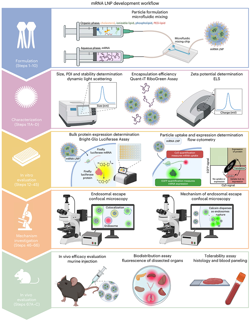
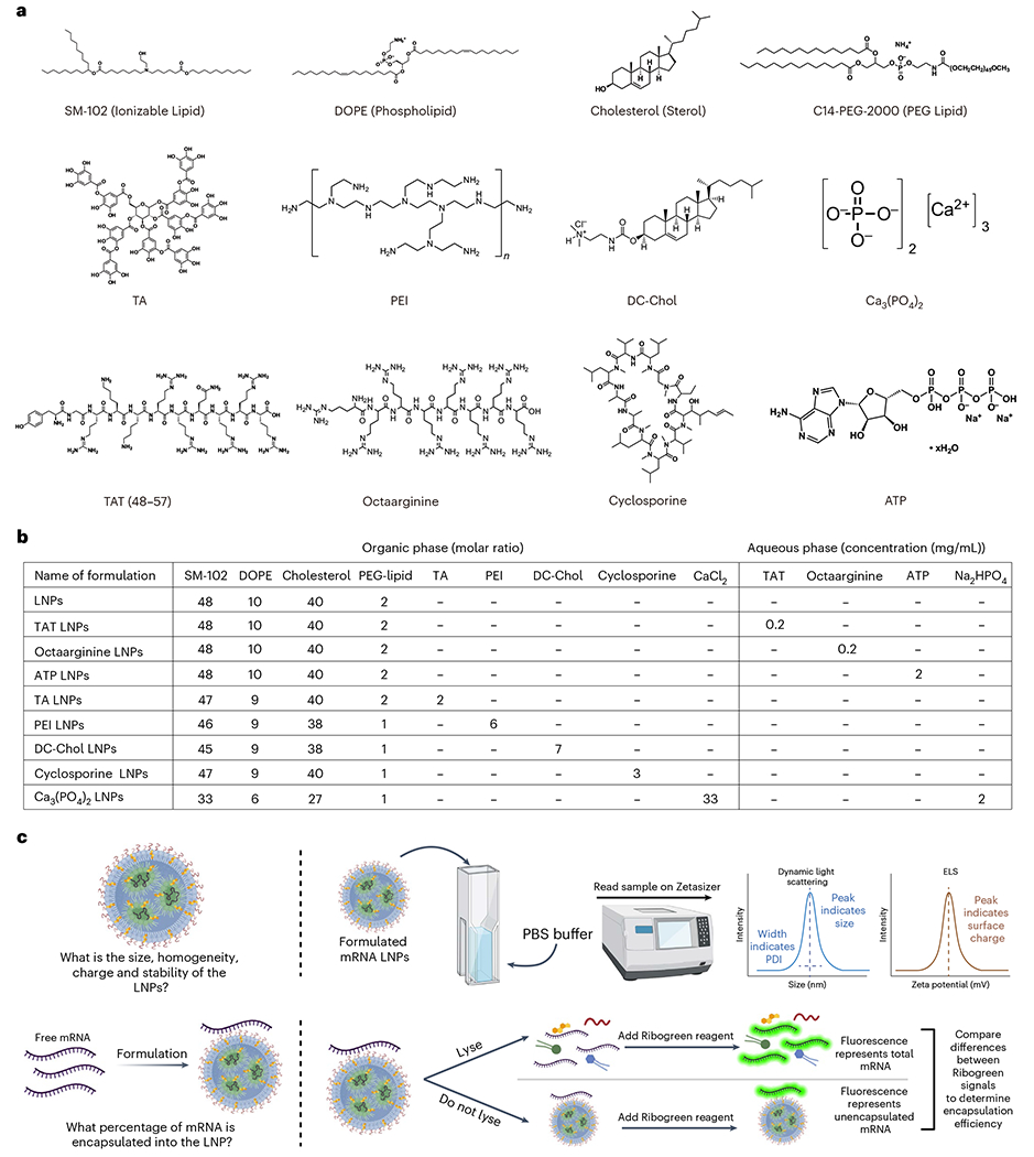
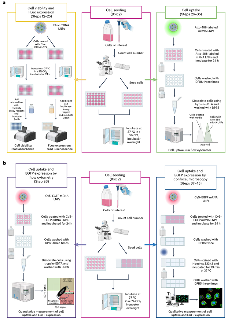
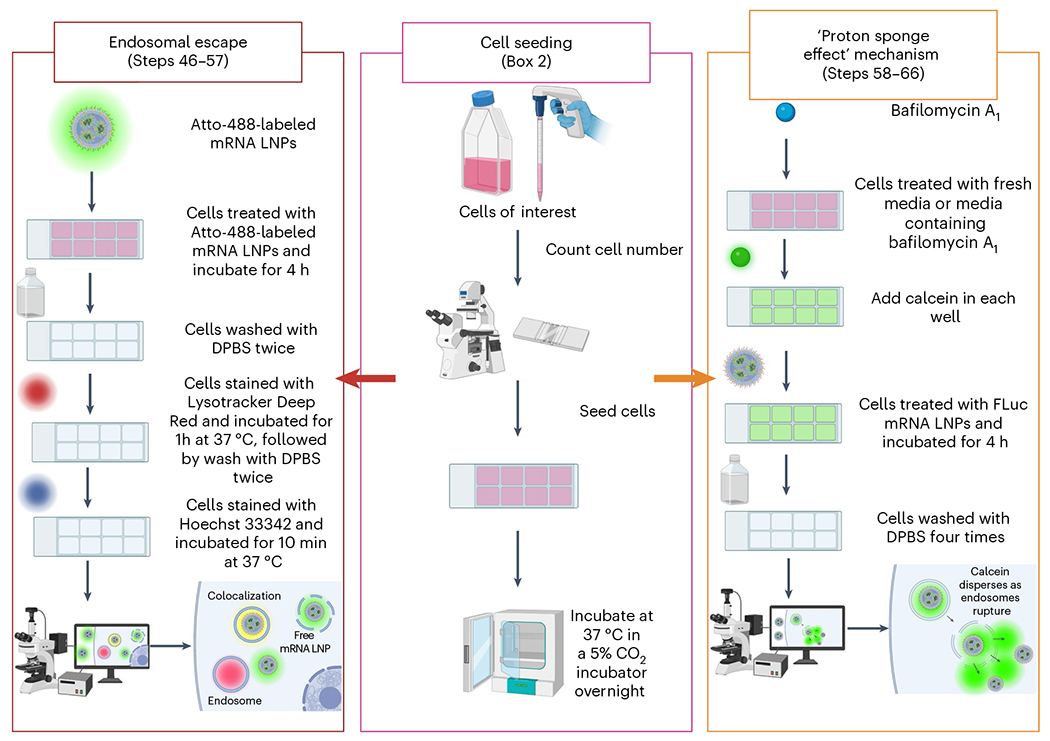
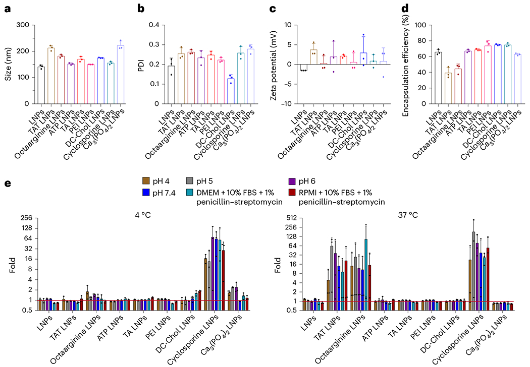
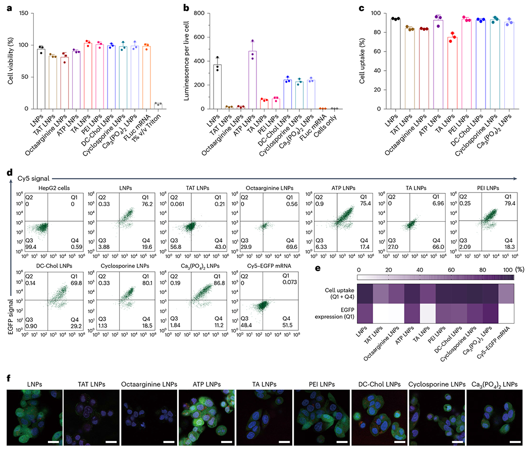
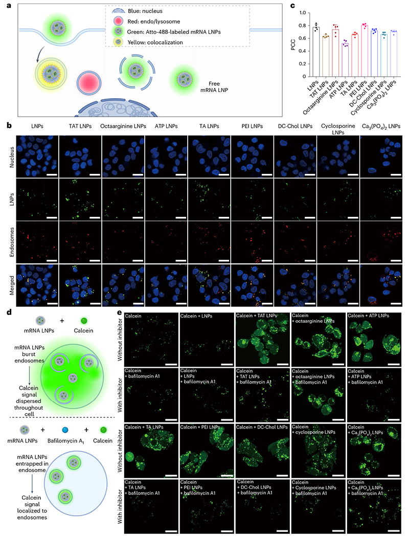
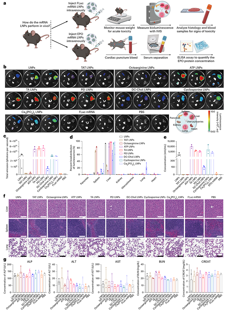
mRNA-based therapies have emerged as a cutting-edge approach for diverse therapeutic applications. However, substantial barriers exist that hinder scientists from entering this research field, including the technical complexity and multiple potential workflows available for formulating and evaluating mRNA lipid nanoparticles (LNPs). Here we present an easy-to-follow and step-by-step guide for mRNA LNP formulation, characterization and in vitro and in vivo evaluation that could lower these barriers, facilitating entry for scientists in academia, industry and clinical settings into this research space. In this protocol, we detail steps for formulating representative mRNA LNPs (0.5 d) and characterizing key parameters (1-6 d) such as size, polydispersity index, zeta potential, mRNA concentration, mRNA encapsulation efficiency and stability. Then, we describe in vitro evaluations (3-6 d), such as protein expression, cell uptake and mechanism investigations (3-5 d), including endosomal escape, as well as in vivo delivery evaluation (2-3 d) encompassing intracellular and secreted protein expression levels, biodistribution and additional tolerability studies (1-2 weeks). Unlike some alternative protocols that may focus on discrete aspects of the workflow-such as formulation, characterization or evaluation-our protocol instead aims to integrate each of these aspects into a simplified, singular workflow applicable across multiple types of mRNA LNP formulations. In describing these procedures, we wish to disseminate one potential workflow for mRNA LNP production and evaluation, with the ultimate goal of furthering innovation, collaboration and the translational advancement of mRNA LNPs.


Conflict of interest statement

Competing interests: The University of North Carolina at Chapel Hill has filed some patents on data related to this work, with the authors named as inventors.

Figures

Fig. 1 |
Fig. 1 |. Overview of the protocol for formulating and evaluating mRNA LNPs.
mRNA LNPs are prepared using a microfluidic mixing approach (Steps 1–10). The key parameters of the obtained mRNA LNPs are then characterized including the size, PDI, zeta potentials, mRNA encapsulation efficiency and stability (Steps 11A–D). The FLuc protein expression and cell uptake are measured using Bright-Glo Luciferase Assay and flow cytometry, respectively (Steps 12–45). The endosomal escape and the mechanism driving LNP release from endosomes is quantified by confocal microscopy (Steps 46–66). The in vivo evaluation of mRNA LNPs is performed by IV injection of mRNA LNPs into mice. Biodistribution, protein expression levels and tolerability assays can be performed to evaluate the mRNA LNPs (Steps 67A–C).
Fig. 2 |
Fig. 2 |. Considerations for the formulation and characterization of mRNA LNPs.
a, The chemical structures of representative molecular excipients that can optionally be used within mRNA LNPs in this study. b, The molar composition ratios for the formulation of mRNA LNPs. c, A schematic illustration of the characterization of the size, PDI, zeta potential and mRNA encapsulation efficiency of mRNA LNPs.
Fig. 3 |
Fig. 3 |. Outline of in vitro evaluation methods for mRNA LNPs.
a, Schematic illustrations of FLuc expression and cell viability of FLuc mRNA LNPs using the Bright-Glo Luciferase Assay and alamarBlue assay, respectively, measured with a microplate reader and a schematic illustration of the cell uptake of Atto-488-labeled mRNA LNPs using flow cytometry. b, Schematic illustrations of quantitative measurement of cell uptake and protein expression using Cy5–EGFP mRNA LNPs by flow cytometry and a qualitative measurement of cell uptake and protein expression using Cy5–EGFP mRNA LNPs by confocal microscopy.
Fig. 4 |
Fig. 4 |. Outline of mechanism investigation methods for mRNA LNPs.
Schematic illustrations of endosomal escape and ‘proton sponge effect’ (one of the mechanisms driving mRNA LNP release from endosomes) assays using confocal microscopy.
Fig. 5 |
Fig. 5 |. LNP formulation and characterization methods.
ad, size/diameter (a), PDI (b), zeta potential (c) and mRNA encapsulation efficiency (d) of mRNA LNPs. e, The stability of different mRNA LNPs after incubation in pH 4, 5, 6 or 7.4, or DMEM + 10% FBS + 1% penicillin–streptomycin or RPMI + 10% FBS + 1% penicillin–streptomycin solutions at either 4 °C or 37 °C for 5 d. The stability was indicated by the size change ratio of day 5 relative to day 0. The horizontal red line represents a size change ratio of 1, indicating no size change after 5 d of incubation and, thus, demonstrating the mRNA LNPs are stable under these conditions (the data presented in ae are shown as mean ± standard deviation, n = 3). Source data are provided in Supplementary Information.
Fig. 6 |
Fig. 6 |. In vitro evaluation methods of mRNA LNPs.
ac, Cell viability (a), FLuc expression (b) and cell uptake (c) of mRNA LNPs; HepG2 cells were treated with mRNA LNPs for 24 h at 37 °C. d, Representative data of EGFP signals versus Cy5 signals on HepG2 cells incubated in the absence (control) or presence of mRNA LNPs for 24 h at 37 °C. Q1 represents the EGFP(+) and Cy5(+) signal (mRNA LNP uptake and EGFP expression), Q2 represents the EGFP(+) and Cy5(−) signal (this is an empty gate, which serves as a control in which we should not see cells if the assay is working correctly), Q3 represents the EGFP(−) and Cy5(−) signal (no mRNA LNPs and no EGFP expression), Q4 represents the EGFP(−) and Cy5(+) signal (mRNA LNP uptake and no EGFP expression). e, A heat map summary of cell uptake and EGFP expression of HepG2 cells treated with Cy5–EGFP mRNA LNPs for 24 h at 37 °C (the data presented in ac and e are shown as the mean ± standard deviation, n = 3) f, Representative confocal images of HepG2 cells treated with Cy5–EGFP mRNA LNPs for 24 h at 37 °C. Scale bars, 20 μm. A statistical analysis of the data in Fig. 6c is shown in Supplementary Fig. 13a. The gating of the data in Fig. 6d is shown in Supplementary Fig. 4. Source data are provided in Supplementary Information.
Fig. 7 |
Fig. 7 |. Mechanism investigation methods.
a, A schematic illustration of endosomal escape studies using a lysotracker confocal imaging assay. b, Representative confocal images of HepG2 cells treated with Atto-488-labeled mRNA LNPs (green). The endosomes (red) are stained with Lysotracker Deep Red. Nuclei (blue) are stained with Hoechst 33342. Scale bars, 20 μm. c, A PCC analysis of Atto-488-labeled mRNA LNPs treated with HepG2 cells (the data are presented as the mean ± standard deviation, and five representative cell images (>20 cells) were used to calculate the PCC value). d, A schematic illustration of endosomal escape mechanism studies using a calcein leakage confocal imaging assay with bafilomycin A1 to test the ‘proton sponge effect’. e, Representative confocal images of HepG2 cells incubated with calcein or calcein and mRNA LNPs in the absence (top) and presence (bottom) of inhibitor bafilomycin A1 for 4 h at 37 °C. Scale bars, 20 μm. A statistical analysis of the data in Fig. 7c is shown in Supplementary Fig. 13b. Source data are provided in Supplementary Information.
Fig. 8 |
Fig. 8 |. In vivo evaluation methods of mRNA LNPs.
a, mRNA LNPs at a dose of 0.65 mg/kg total mRNA are intravenously injected into mice (6–8 weeks old, weight 18–21 g). After 24 h, the organs were imaged by IVIS imaging. The blood was collected to run the blood toxicity. The organs were collected to run histology. The weight was monitored to quantify the acute toxicity. b, Bioluminescence images of various organs ex vivo for each group 24 hours after IV administration of mRNA LNPs. The mice injected with naked FLuc mRNA and only PBS served as controls. c,d, Total luminescence (c) and associated percent of bioluminescence (d) of mRNA LNPs across various organs including the pancreas, spleen, liver, kidneys, uterus/ovaries, lung and heart. e, Human EPO concentrations after the injection of EPO mRNA LNPs for 24 h. The mice injected with naked EPO mRNA and only PBS served as controls. The concentration of human EPO was characterized by human EPO ELISA kits following the manufacturer’s protocol. f, Representative histology images of the liver, spleen and lungs of mice after treatment with mRNA LNPs via IV injection. Scale bars, 100 μm. g, ALP, ALT, AST, BUN and creatinine (CREAT) blood testing results after the IV injection of mRNA LNPs. A statistical analysis of data in Fig. 8c,e is shown in Supplementary Fig. 13c,d (The data presented in ce and g are shown as the mean ± standard deviation, n = 3). Source data are provided in Supplementary Information.

Similar articles

Cited by

References

    1. Kon E, Ad-El N, Hazan-Halevy I, Stotsky-Oterin L & Peer D Targeting cancer with mRNA-lipid nanoparticles: key considerations and future prospects. Nat. Rev. Clin. Oncol 20, 739–754 (2023). - PubMed
    1. Li F et al. mRNA lipid nanoparticle-mediated pyroptosis sensitizes immunologically cold tumors to checkpoint immunotherapy. Nat. Commun 14, 4223 (2023). - PMC - PubMed
    1. Neill B, Romero AR & Fenton OS Advances in nonviral mRNA delivery materials and their application as vaccines for melanoma therapy. ACS Appl. Bio Mater 7, 4894–4913 (2024). - PMC - PubMed
    1. Wang C, Zhang Y & Dong Y Lipid nanoparticle–mRNA formulations for therapeutic applications. Acc. Chem. Res 54, 4283–4293 (2021). - PMC - PubMed
    1. Jeong M, Lee Y, Park J, Jung H & Lee H Lipid nanoparticles (LNPs) for in vivo RNA delivery and their breakthrough technology for future applications. Adv. Drug Deliv. Rev 200, 114990 (2023). - PubMed

Key references

    1. Ma Y & Fenton OSJ Am. Chem. Soc 145, 19800–19811 (2023): 10.1021/jacs.3c05574 - DOI - PubMed
    1. Ma Y & Fenton OSJ Am. Chem. Soc 145, 11375–11386 (2023): 10.1021/jacs.3c02584 - DOI - PubMed
    1. Ma Y & Fenton OS Adv. Therap 6, 2200305 (2023): 10.1002/adtp.202200305 - DOI

LinkOut - more resources