Review
. 2025 Mar 11;23(1):131.
doi: 10.1186/s12964-025-02090-6.

Dys-regulated phosphatidylserine externalization as a cell intrinsic immune escape mechanism in cancer

Affiliations
Review

Dys-regulated phosphatidylserine externalization as a cell intrinsic immune escape mechanism in cancer

Rachael Pulica et al. Cell Commun Signal. .

Abstract

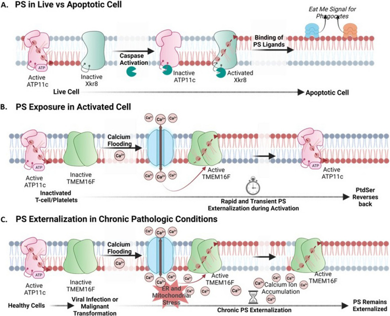
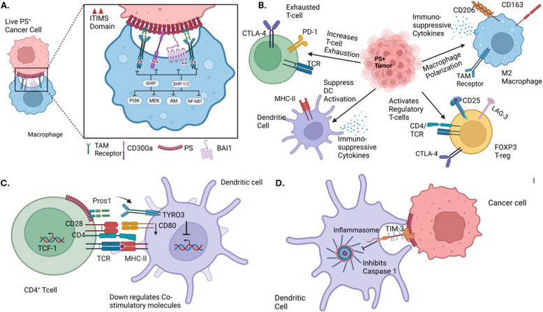
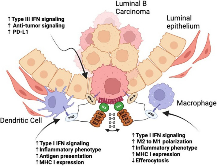
The negatively charged aminophospholipid, phosphatidylserine (PS), is typically restricted to the inner leaflet of the plasma membrane under normal, healthy physiological conditions. PS is irreversibly externalized during apoptosis, where it serves as a signal for elimination by efferocytosis. PS is also reversibly and transiently externalized during cell activation such as platelet and immune cell activation. These events associated with physiological PS externalization are tightly controlled by the regulated activation of flippases and scramblases. Indeed, improper regulation of PS externalization results in thrombotic diseases such as Scott Syndrome, a defect in coagulation and thrombin production, and in the case of efferocytosis, can result in autoimmunity such as systemic lupus erythematosus (SLE) when PS-mediated apoptosis and efferocytosis fails. The physiological regulation of PS is also perturbed in cancer and during viral infection, whereby PS becomes persistently exposed on the surface of such stressed and diseased cells, which can lead to chronic thrombosis and chronic immune evasion. In this review, we summarize evidence for the dysregulation of PS with a main focus on cancer biology and the pathogenic mechanisms for immune evasion and signaling by PS, as well as the discussion of new therapeutic strategies aimed to target externalized PS. We posit that chronic PS externalization is a universal and agnostic marker for diseased tissues, and in cancer, likely reflects a cell intrinsic form of immune escape. The continued development of new therapeutic strategies for targeting PS also provides rationale for their co-utility as adjuvants and with immune checkpoint therapeutics.

Keywords: Immune escape; P4 ATPase; PS receptors; Phosphatidylserine; Scramblases.


Conflict of interest statement

Declarations. Competing interests: SVK and RBB are co-founders of a biotech company called Targeron Therapeutics that aim to develop PS-targeting biologicals. Other authors have nothing to declare.

Figures

Fig. 1
Fig. 1
Intracellular and extracellular pools of PS. The proteomes of cells internalizing versus externalizing PS are unique from one another. When PS is on the inner side of the PM, it binds signaling proteins and cytoskeletal proteins. In contrast, on the exterior of the PM, coagulation factors and immunoregulatory molecules remain unbound. Upon PS externalization, the inner membrane becomes the released proteome, meaning that these signaling and cytoskeletal proteins are released from the PM. Meanwhile, on the external side of the cell, PS binds up coagulation factors and immunoregulatory molecules, resulting in the induction of their given pathways
Fig. 2
Fig. 2
PS synthesis at the mitochondria-associated membranes (MAMs) and transport to the plasma membrane (PM). (1) PS is first synthesized via the PSS1/PSS2-mediated conversion of PC and PE to PS at the MAMs. (2) PS is then transported to the outer mitochondrial membrane and inner PM by lipid transfer proteins (e.g. ORP5/8). (3) Unrestricted lateral movement of PS then shifts it across the PM. (4) Once integrated in the PM, PS is externalized by scramblases and internalized by P4-ATPases.​ These arrangements are expected to restrict PS binding proteins that are destined for secretion to become trapped by intracellular pools for PS
Fig. 3
Fig. 3
Chronic and reversible PS externalization by scramblases. (A) In live cells, PS is internalized due to the activity of P4-ATPase ATP11C and the inactivity of scramblase Xkr8. During apoptosis, caspases cleave ATP11C and Xkr8 at their caspase cleavage site, rendering them inactive and activated respectively. This leads to the irreversible externalization of PS and the binding of PS ligands, resulting in “eat-me signals” for phagocytosis. (B) Inactivated T cells and platelets internalize PS due to active ATP11C and inactive TMEM16F. Upon calcium influx into the cytosol (activation), TMEM16F forms an active dimer and externalizes PS rapidly and temporarily. Active ATP11C can then internalize PS again upon restoration of calcium homeostasis. Healthy cells typically possess active ATP11C and inactive TMEM16F, maintaining PS on the intracellular leaflet (C). Upon pathophysiological conditions of viral infection or malignant progression, calcium levels in the cytosol become constitutively elevated leading to constitutive TMEM16F activation and chronic PS externalization
Fig. 4
Fig. 4
PS externalization in live versus apoptotic cells. Viable cells (left) have PS externalized (shown by the binding of PS-targeting antibody, 11.31) by scramblase TMEM16F in a punctate, localized manner. This is then brought back in due to the hydrolysis of ATP to ADP and the resulting activities of ATP11C. In contrast, PS exposed on apoptotic cells (right) due to caspase cleavage of ATP11C and scramblase Xkr8 show PS externalized throughout the entirety of the PM in a less localized and more uniform manner [115]
Fig. 5
Fig. 5
Immunomodulatory effects of PS externalization in the TME. A. Live PS-expressing cancer cells engage PS-binding TAM, CD300a, and BAI1 receptors on macrophages. These receptors contain ITIM or ITSM motifs, which recruit SHP1/2 or SHIP phosphatases to inhibit downstream pro-inflammatory signaling pathways, including NF-κB, Akt, MEK, and PI3K, promoting immune suppression. B. Chronic PS exposure in the TME sustains immune suppression by polarizing macrophages to M2 macrophages, converting DCs toward resolving phenotype, increasing T cell exhaustion, and activating Regulatory T cells. C. Activated T cells secrete Pros1, a ligand for Tyro3 expressed on dendritic cells, dampening antigen presentation and limiting co-stimulatory molecule expression. D. PS externalization on cancer cells can also inhibit the NLRP3 inflammasome via TIM-3 receptor engagement, dampening inflammatory signaling (see text for details)
Fig. 6
Fig. 6
Efferocytosis of apoptotic cells, lysosomal degradation, and efferocyte reprograming. (Panel 1) Externalized PS is an emblematic signal for both recognition and trafficking of apoptotic cells to the phagolysosome. (Panel 2) Mertk is proposed to act as a “trojan horse” to traffic apoptotic cells to intracellular itineraries and the lysosome. (Panel 3) Phagolysosomes then degrade and recycle components of the phagolysosome to the cytosol and other intracellular compartments for phenotypic reprogramming. (Panel 4) Macrophage phenotype is now an M2c-like state wherein efferocytosis can be furthered and a pro-tumoral environment can be achieved. These M2c macrophages are thought to promote tumor progression and metastasis while suppressing inflammation
Fig. 7
Fig. 7
Mechanisms for chronic PS externalization in the TME. Several mechanisms have been proposed for the tonic sustained PS externalization in the tumor microenvironment that include scramblase overexpression (A), upregulation/elevated levels of calcium channels (B), hypoxia and ROS (C), increased extracellular ATP (D), oncogenic kinases phosphorylating Xkr8 (E), and mutation of flippases all contribute to the constant PS externalization in the TME that leads to a cold tumor phenotype (F)
Fig. 8
Fig. 8
Strategies to target externalized PS in cancer. PS can be targeted both intrinsically and extrinsically to achieve an antitumoral immune stimulatory outcome. Cell extrinsic therapies (left) include modalities that bind to cell externalized PS as well as modalities to inhibit PS scrambling. They are proposed to reduce or nullify the ability for PS to polarize the TME (or reduce the quantity of PS overall) as well as deliver therapeutics with PS as the homing beacon to the TME. Cell intrinsic methods (right) may become internalized within the cell upon PS binding to deliver intracellular therapeutics
Fig. 9
Fig. 9
Fusion of PS-targeting domains with dual interferons provides a novel immune stimulating payload. Gas6-IFN-β IFN-λ provides a PS targeting modality combined with dual acting IFNs as an immunogenic payload. The IFNβ is expected then bind any nucleated immune cells in the TME, while the IFNλ binds epithelial cells, including the PS exposed on the tumor cells. IFN signaling will then result in an inflammatory, hot tumor phenotype and reduced efferocytosis
Fig. 10
Fig. 10
PS externalization on the surface of enveloped viruses and convergent evolution. While viral surfaces display a multitude of different cell surface proteins on their surfaces most, if not all, enveloped viruses share a common characteristic of PS externalized on the surface called apoptotic mimicry. Conceptually, apoptotic mimicry supports the generalized idea that PS represents a universal immunosuppressive mechanism to divert host immunity and that PS is also a promising option to target for antiviral therapeutics

References

    1. Lenard J, Rothman JE. Transbilayer distribution and movement of cholesterol and phospholipid in the membrane of influenza virus. Proc Natl Acad Sci U S A. 1976;73:391–5. - PMC - PubMed
    1. Rothman JE, Lenard J. Membrane asymmetry. Science. 1977;195:743–53. - PubMed
    1. Bergelson LD, Barsukov LI. Topological asymmetry of phospholipids in membranes. Science. 1977;197:224–30. - PubMed
    1. Leventis PA, Grinstein S. The distribution and function of phosphatidylserine in cellular membranes. Annu Rev Biophys. 2010;39:407–27. - PubMed
    1. Sigal CT, Zhou W, Buser CA, McLaughlin S, Resh MD. Amino-terminal basic residues of Src mediate membrane binding through electrostatic interaction with acidic phospholipids. Proc Natl Acad Sci U S A. 1994;91:12253–7. - PMC - PubMed

Substances

LinkOut - more resources