. 2025 Mar 11;23(1):200.
doi: 10.1186/s12951-025-03293-2.

Young fibroblast-derived migrasomes alleviate keratinocyte senescence and enhance wound healing in aged skin

Affiliations

Young fibroblast-derived migrasomes alleviate keratinocyte senescence and enhance wound healing in aged skin

Hanlin Tu et al. J Nanobiotechnology. .

Abstract

Background: Alterations in intercellular communication driven by cellular senescence constitute an important factor in skin aging. Migrasome, a newly discovered vesicular organelle, efficiently participates in intercellular communication; however, the relationship between cellular senescence and migrasomes remains unreported.

Objective: This study aims to explore the possible relationship between cellular senescence and migrasomes formation, and investigate the effects of young fibroblast-derived migrasomes on senescent keratinocytes and wound healing in aged skin.

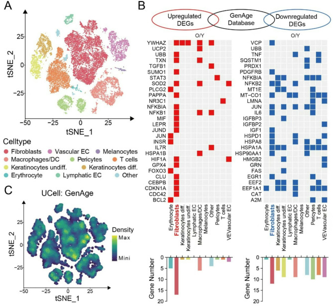
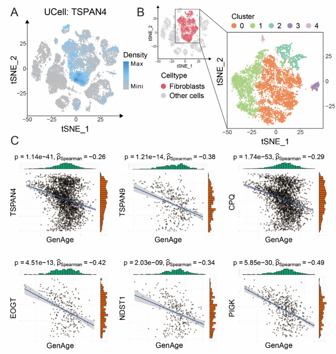
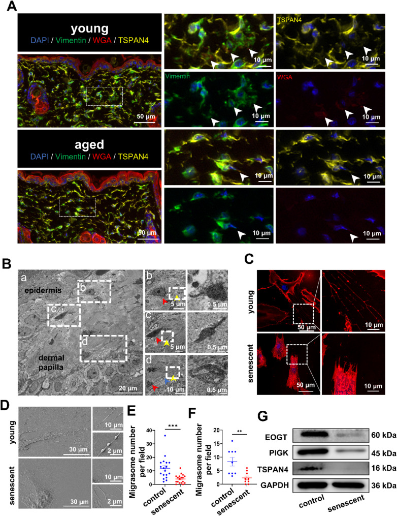
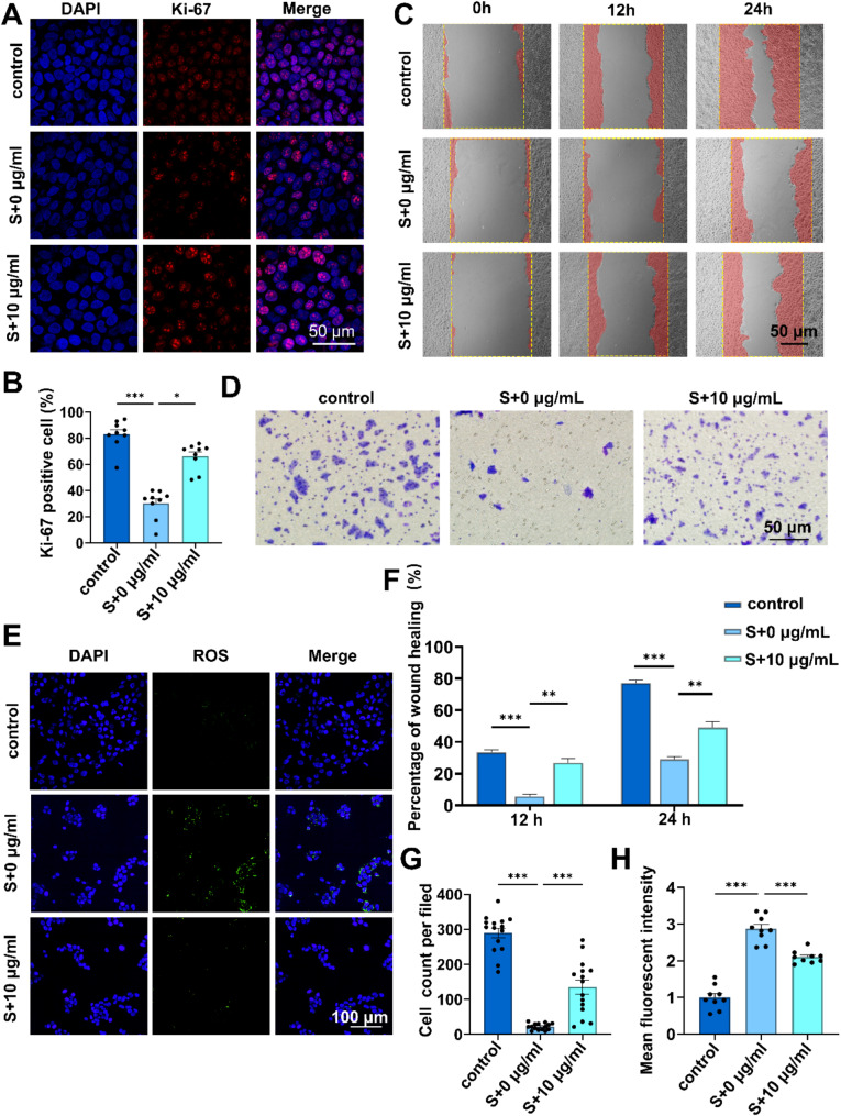
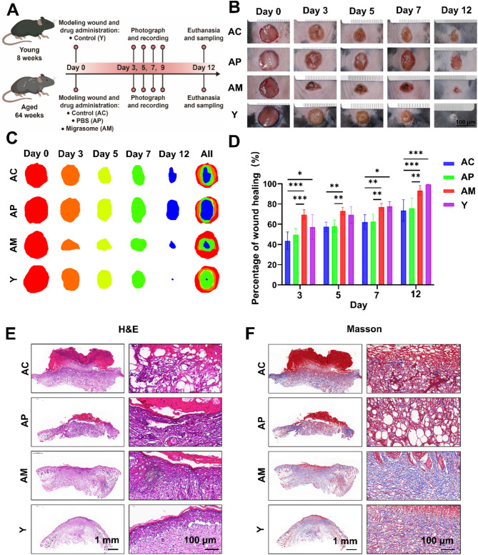
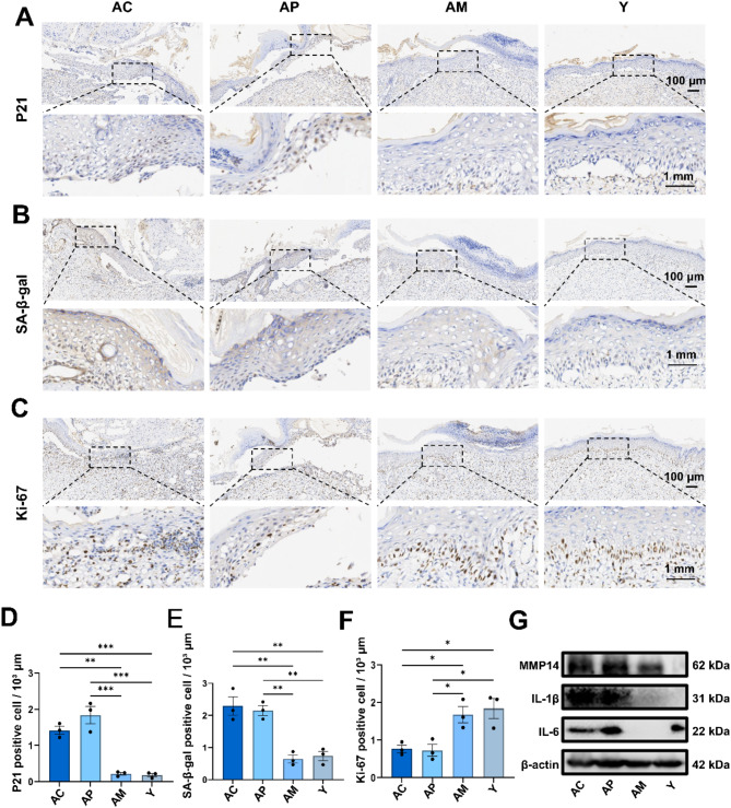
Result: Single-cell RNA sequencing (scRNA-seq) data analysis revealed that fibroblasts exhibited the highest level of transcriptional variability during skin aging, and the degree of fibroblast senescence negatively correlated with the expression level of migrasome-associated markers. Further multiplex Immunohistochemistry (mIHC) results suggested that younger mouse skin contained more migrasomes than older mouse skin. Transmission electron microscopy (TEM) observations demonstrated abundant migrasomes in the skin from young individuals. In vitro experiments indicated that young fibroblasts produced significantly more migrasomes than senescent fibroblasts, as confirmed by wheat germ agglutinin (WGA) staining and scanning electron microscopy (SEM). Importantly, purified migrasomes from young fibroblasts were found to reduce the expression of senescence-associated markers in HaCaT cells. In vivo, using a wound healing model in naturally aged mice, we observed that migrasomes derived from young fibroblasts not only accelerated wound healing but also reduced senescence-associated marker expression in the skin.

Conclusion: Migrasomes formation ability reduced during skin aging progress, and young fibroblast-derived migrasomes rejuvenated senescent keratinocytes and promoted wound healing in aged skin. These findings offer new ideas for alleviating skin aging and enhancing wound healing in aged skin.

Keywords: Aging; Fibroblast; Migrasomes; Senescence; Skin wound healing.


Conflict of interest statement

Declarations. Competing interests: The authors declare no competing interests.

Figures

Fig. 1
Fig. 1
Single-cell RNA sequencing analysis reveals the role of fibroblasts in aging skin A. t-Distributed Stochastic Neighbor Embedding (t-SNE) plot depicting single-cell transcriptomes from whole human skin (n = 5). Each dot represents a single cell (n = 25,031). Cells are colored by types and annotated to the right B. Heatmap showing genes shared between aging-related genes in the GenAge database and aging-associated DEGs for each skin cell type. Only the ensembles of shared genes are shown, and the number statistics are shown below the heatmap C. Mapping density plots of t-SNE showing scoring results from GenAge databases (see Methods for details). Yellow color indicates maximum gene expression and dark blue color indicates low or no gene expression
Fig. 2
Fig. 2
ScRNA-seq suggests a potential relationship between aging and migrasomes formation A. Mapping density plot of t-SNE showing the scoring result for the migrasome-associated gene TSPAN4. Dark blue color indicates maximum gene expression and grey color indicates low or no gene expression B. t-SNE diagram illustrated the cell clusters of fibroblasts C. Scatterplots showing the correlation between the six migrasome-associated genes (TSPAN4, TSPAN9, CPQ, EOGT, NDST1 and PIGK) and aging-related genes in the GenAge database respectively (see Methods for details). The trend lines represent the overall trend of all the scatter points in the corresponding plots, and the corresponding p-value and Spearman’s correlation coefficient are shown at the top of the plots
Fig. 3
Fig. 3
Senescent fibroblasts significantly impair migrasomes formation A. mIHC of skin from young and aged mice. Skin sections from three 8-wFeek-old young mice and three 64-week-old aged mice were subjected to vimentin, TSPAN4, and WGA staining. Vimentin (green) identifies fibroblasts, while the colocalization of TSPAN4 (yellow) and WGA (red) denotes migrasomes. White arrows denote colocalization of Vimentin, TSPAN4 and WGA. Representative images are presented B. TEM of skin sample from young individuals. (a) Shows the dermal papilla structure; (b, c, d) present magnified views of migrasome-like structures. Yellow arrows denote migrasome-like structures, blue arrows indicate retraction fiber-like structures, and red arrows highlight potential migrasome precursor cells C. WGA staining images of young BJ cells and H2O2-induced senescent BJ cells, with magnified views D. SEM images of young BJ cells and H2O2-induced senescent BJ cells, with magnified views E. Quantification of migrasome numbers in young BJ cells and H2O2-induced senescent BJ cells using WGA staining. The experiment was independently repeated three times, with six random fields analyzed per group. Data are presented as mean ± SEM with significance (by normality and lognormality test followed by Mann-Whitney test) F. Quantification of migrasome numbers in young BJ cells and H2O2-induced senescent BJ cells was performed using scanning electron microscopy. The experiment was independently repeated three times, with three random fields analyzed per group. Data are presented as mean ± SEM with significance (by normality and lognormality test followed by Welch’s t test) G. Expression levels of migrasome markers in young BJ cells and H₂O₂-induced senescent BJ cells analyzed by Western blot
Fig. 4
Fig. 4
Young fibroblast-derived migrasomes alleviate senescence in HaCaT in vitro A. Figure of the purification procedure for young fibroblast-derived migrasomes B. Representative TEM images of negative staining for purified young fibroblast-derived migrasomes C. Representative images of western blot showing the expression of migrasome-related markers in purified young fibroblast-derived migrasomes D. The internalization of migrasomes by senescent HaCaT cells. The migrasomes are specifically labeled with WGA and appear as red puncta, and phalloidin staining in green E. Representative images of SA-β-gal staining in H₂O₂-induced senescent HaCaT cells treated with different concentrations of young fibroblast-derived migrasomes F. Representative Western Blot images showing the expression of senescence markers p16 and p21 in H₂O₂-induced senescent HaCaT cells treated with different concentrations of young fibroblast-derived migrasomes n G. Quantification of SA-β-gal positive cells in H₂O₂-induced senescent HaCaT cells treated with different concentrations of migrasomes for 48 h. Experiments were repeated independently three times. Data were collected by randomly photographing 3 fields per group. Statistical analysis was performed using one-way ANOVA. Error bars indicate the mean ± SEM H and I. Quantitative Western Blot analysis showing the expression of senescence markers p16 and p21 in H₂O₂-induced senescent HaCaT cells after 48 h of stimulation with various concentrations of migrasomes. Experiments were conducted independently three times. Data were analyzed using one-way ANOVA. Error bars indicate the mean ± SEM
Fig. 5
Fig. 5
Young fibroblast-derived migrasomes promote senescent HaCaT cells functions. HaCaT cells were co-cultured with or without the young fibroblast-derived migrasomes (10 µg/ml) following treatment with H₂O₂ (600 µmol/L) for 4 h A. Representative images of Ki-67 (red) and DAPI (blue) immunofluorescence staining in HaCaT cells B. Quantification of Ki-67 immunofluorescence staining in HaCaT cells. Experiments were conducted independently three times. Data are presented as mean ± SD with significance determined by normality and lognormality tests followed by the Kruskal-Wallis test C. Representative images of the scratch assay at 12 h and 24 h D. Representative images of the transwell migration assay in HaCaT cells E. Representative images of ROS (red) and DAPI (blue) staining in HaCaT cells F. Quantification of the scratch assay at 12 h and 24 h. Experiments were conducted independently three times. Data were analyzed using one-way ANOVA. Error bars represent the mean ± SEM G. Quantification of transwell migration in HaCaT cells. Experiments were performed independently three times. Data are presented as mean ± SEM. Statistical significance was assessed using normality and lognormality tests, followed by the Brown-Forsythe test and Welch’s ANOVA H. Quantification of Ki-67 immunofluorescence staining in HaCaT cells. Experiments were conducted independently three times. Data were analyzed using one-way ANOVA. Error bars represent the mean ± SEM
Fig. 6
Fig. 6
In vivo effects of young fibroblast-derived migrasomes on wound healing in aging mouse skin A. Schematic of the experimental design for the mouse wound healing model B and C. Representative images of skin wounds in young and aging C57BL/6 mice treated with PBS or young fibroblast-derived migrasomes (100 µg) at days 0, 3, 6, 9, and 12 post-surgeries. The mice were randomly assigned to four groups: Young Mouse Group (Y), Aged Mouse Control Group (AC), Aged Mouse PBS-Injected Group (AP), and Aged Mouse Migrasome-Injected Group (AM). The young mouse group consists of 3 mice, and the aging mouse groups consist of 5 mice each.D. Quantification of wound area during the healing process. Data are presented as mean ± SD. Young mice (n = 3) and aging mice (n = 5) were analyzed. Statistical significance was assessed using two-way ANOVA. Error bars represent the mean ± SD E. Representative images of skin wounds stained with hematoxylin and eosin (H&E) after 12 days of treatment F. Representative images of skin wounds stained with Masson’s trichrome after 12 days of treatment
Fig. 7
Fig. 7
In vivo effects of young fibroblast-derived migrasomes on reducing senescent cells and SASP A. Representative immunohistochemical staining images for p21 B. Representative immunohistochemical staining images for SA-β-gal C. Representative immunohistochemical staining images for Ki-67 D. Quantification of immunohistochemical staining images for p21. Experiments were conducted independently three times. Data were analyzed using one-way ANOVA. Error bars represent the mean ± SEM E. Quantification of immunohistochemical staining images for SA-β-gal. Experiments were conducted independently three times. Data were analyzed using one-way ANOVA. Error bars represent the mean ± SEM F. Quantification of immunohistochemical staining images for Ki-67. Experiments were conducted independently three times. Data were analyzed using one-way ANOVA. Error bars represent the mean ± SEM G. Representative Western Blot images showing the expression of IL-1β, IL-6 and MMP14

Similar articles

References

    1. Ghadially R, Brown BE, Sequeira-Martin SM, Feingold KR, Elias PM. The aged epidermal permeability barrier. Structural, functional, and lipid biochemical abnormalities in humans and a senescent murine model. J Clin Invest. 1995;95:2281–90. - PMC - PubMed
    1. Velarde MC, Demaria M. Targeting senescent cells: possible implications for delaying skin aging: A Mini-Review. Gerontology. 2016;62:513–8. - PubMed
    1. Sgonc R, Gruber J. Age-related aspects of cutaneous wound healing: a mini-review. Gerontology. 2013;59:159–64. - PubMed
    1. Dimri GP, Lee X, Basile G, Acosta M, Scott G, Roskelley C, Medrano EE, Linskens M, Rubelj I, Pereira-Smith O, et al. A biomarker that identifies senescent human cells in culture and in aging skin in vivo. Proc Natl Acad Sci U S A. 1995;92:9363–7. - PMC - PubMed
    1. Wang B, Han J, Elisseeff JH, Demaria M. The senescence-associated secretory phenotype and its physiological and pathological implications. Nat Rev Mol Cell Biol. 2024;25:958–78. - PubMed

LinkOut - more resources