Review
. 2025 Jan-Dec:21:17448069251327840.
doi: 10.1177/17448069251327840. Epub 2025 Mar 11.

Exploring novel non-opioid pathways and therapeutics for pain modulation

Affiliations
Review

Exploring novel non-opioid pathways and therapeutics for pain modulation

Isabella Alessi et al. Mol Pain. 2025 Jan-Dec.

Abstract

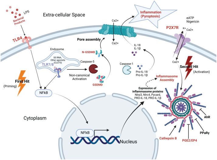
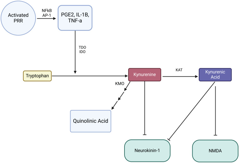
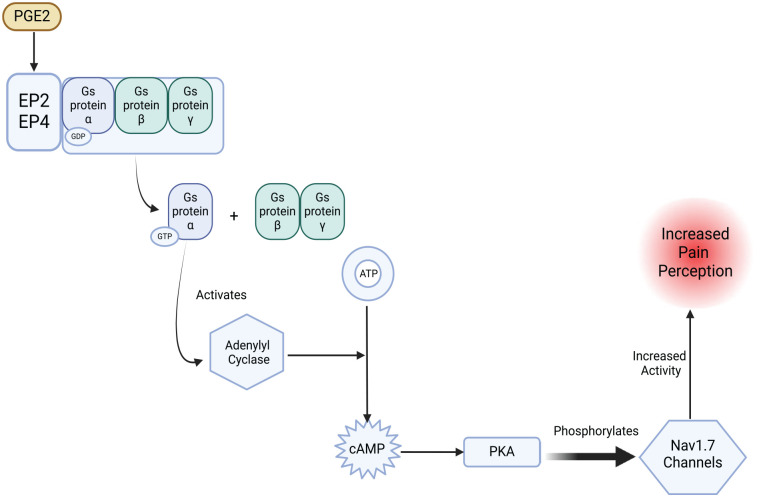
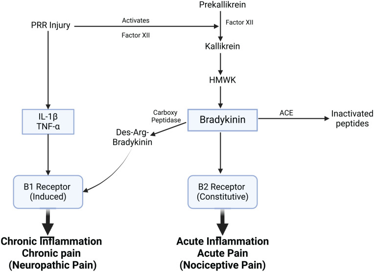
The opioid crisis has highlighted the urgent need for alternative pain management strategies. This review explores novel non-opioid targets and pathways involved in pain modulation, highlighting advancements in understanding and therapeutic potential. Pain, a multifaceted phenomenon with nociceptive, neuropathic, and inflammatory components, involves intricate molecular signaling cascades. Key pathways reviewed include voltage-gated sodium channels (Nav1.7, Nav1.8, Nav1.9), inflammasome complexes (NLRP3), the kynurenine pathway, prostaglandins, and bradykinin-mediated signaling. Emerging therapeutics such as selective Nav channel blockers, NLRP3 inhibitors, kynurenine pathway modulators, EP receptor antagonists, and bradykinin receptor antagonists offer promising alternatives to opioids. Despite challenges in clinical translation, these developments signal a paradigm shift in pain management, with precision-focused therapies poised to address unmet needs. This review emphasizes the importance of integrating molecular insights into the development of safer, more effective analgesics, setting the stage for transformative advancements in non-opioid pain relief.

Keywords: Nav; PGE2; Pain; bradykinin; inflammasome; kynurenine; non-opioid.


Conflict of interest statement

Declaration of conflicting interestsThe author(s) declared no potential conflicts of interest with respect to the research, authorship, and/or publication of this article.

Figures

Figure 1.
Figure 1.
The inflammasome pathway in pain modulation. AHR: Aryl hydrocarbon receptor; ASC: Apoptosis-associated speck like protein containing a caspase recruitment domain; LPS: Lipopolysaccharide; NFκB: Nuclear factor kappa B; NLRP3: NOD-like receptor protein 3; NLRc4: NOD-like receptor c4; PPaRγ: Peroxisome proliferator-activated receptor gamma; TLR: Toll-like receptor. Inflammasome activation occurs in a two key step process – priming and activation. Priming occurs through the recognition of bacterial products such as lipopolysaccharide (LPS) via Toll-like receptors (TLRs). This engagement triggers the nuclear factor kappa B (NF-κB) pathway, leading to the transcription of inflammasome-related components, including NOD-like receptor protein 3 (NLRP3), apoptosis-associated speck like protein containing a caspase recruitment domain (ASC), pro-IL-1β, and pro-IL-18. Activation follows a second hit, commonly involving extracellular ATP binding to purinergic P2X7 receptors, resulting in K⁺ efflux and Ca²⁺ influx. These ion fluxes trigger the assembly of the inflammasome complex, including NLRP3, ASC, and caspase-1. Caspase-1 then processes pro-IL-1β and pro-IL-18 into their active forms, IL1β and IL-18, which are released to propagate inflammation. In some instances, the non-canonical pathway is activated via intracellular recognition of LPS or RNA through caspase-5. Caspase-5 or caspase-1 cleave gasdermin D (GSDMD) into its active N-terminal form, which forms membrane pores to induce pyroptosis and amplify cytokine release. Together, these pathways ensure a robust inflammatory response, mediated by the inflammasome complex. This comprehensive figure highlights the molecular interplay between priming (via TLR signaling) and activation (via ATP and P2X7), demonstrating the canonical role of caspase1 (effector component of inflammasome) and the auxiliary contribution of caspase-5 and GSDMD in driving inflammation.
Figure 2.
Figure 2.
Mechanism of the kynurenine pathway in pain modulation. The figure illustrates how inflammatory mediators interact with the kynurenine pathway to modulate pain. Activation of pattern recognition receptors (PRRs) by damage-or pathogen-associated molecular patterns (DAMPs or PAMPs) initiates downstream signaling cascades, such as NF-κB and AP-1 transcription factors, which upregulate pro-inflammatory molecules, including IL-1β, TNF-α, and PGE2. These inflammatory signals stimulate the conversion of tryptophan into kynurenine through enzymes such as indoleamine 2,3- dioxygenase (IDO) and tryptophan 2,3-dioxygenase (TDO). Kynurenine is then metabolized by kynurenine aminotransferase (KAT) into kynurenic acid, which acts on NMDA receptors to inhibit glutamate signaling, thus reducing excitatory neurotransmission and pain perception. (Note: The role of kynurenic acid in directly inhibiting Neurokinin-1 is under further investigation). For more reference on the complete Kynurenine pathway see: Hazrati et al.
Figure 3.
Figure 3.
The role of PGE2 and EP receptor signaling in pain modulation via Nav1.7 channels. The proposed figure illustrates the signaling cascade through which PGE2 modulates pain. When PGE2 binds to prostaglandin receptors EP2 and EP4 on nociceptive neurons, it activates the Gs protein complex associated with these receptors. This binding initiates a cascade, where Gs protein activation leads to the exchange of GDP for GTP on the Gα subunit, which subsequently activates adenylyl cyclase. Adenylyl cyclase converts ATP to cyclic AMP (cAMP), which in turn activates protein kinase A (PKA). PKA phosphorylates Nav1.7 sodium channels, increasing their activity and enhancing pain signaling. This pathway highlights potential therapeutic targets, such as blocking EP receptors or preventing Nav1.7 phosphorylation, to reduce pain perception effectively.
Figure 4.
Figure 4.
Bradykinin pathway and receptor signaling in inflammatory pain modulation. ACE: Angiotensin converting enzyme; HMWK: High molecular wright kininogen; PRR: Pattern recognition receptors. This figure illustrates the complex pathways involved in bradykinin production and its role in pain signaling. PRR or injury activates Factor XII which cleaves prekallikrein to kallikrein. Kallikrein cleaves HMWK to form bradykinin. Bradykinin binds to the B2 (constitutive) receptor and is responsible to acute inflammatory pain. B1 receptor is induced by inflammation. Bradykinin can be catalyzed by carboxypeptidases to form Des-Argbradykinin which binds to the B1 receptor and is associated in chronic inflammatory pain, (neuropathic pain).

Similar articles

Cited by

References

    1. Loeser JD, Treede RD. The Kyoto protocol of IASP basic pain terminology. Pain 2008; 137(3): 473–477. - PubMed
    1. Yam MF, Loh YC, Tan CS, Khadijah Adam S, Abdul Manan N, Basir R. General pathways of pain sensation and the major neurotransmitters involved in pain regulation. Int J Mol Sci 2018; 19(8): 2164. - PMC - PubMed
    1. Oronsky B, Caroen S, Reid T. What exactly is inflammation (and what is it not?). Int J Mol Sci 2022; 23(23): 14905. - PMC - PubMed
    1. Armstrong SA, Herr MJ. Physiology, nociception. Treasure Island, FL: StatPearls Publishing, 2024. - PubMed
    1. Hange N, Poudel S, Ozair S, Paul T, Nambakkam M, Shrestha R, Greye F, Shah S, Raj Adhikari Y, Thapa S, Patel P. Managing chronic neuropathic pain: recent advances and new challenges. Neurol Res Int 2022; 2022: 8336561. - PMC - PubMed

LinkOut - more resources