. 2025 Feb 7;16(5):2190-2201.
doi: 10.1039/d4md00833b. eCollection 2025 May 22.

Site-specific molecular glues for the 14-3-3/Tau pS214 protein-protein interaction via reversible covalent imine tethering

Affiliations

Site-specific molecular glues for the 14-3-3/Tau pS214 protein-protein interaction via reversible covalent imine tethering

Ansgar Oberheide et al. RSC Med Chem. .

Abstract

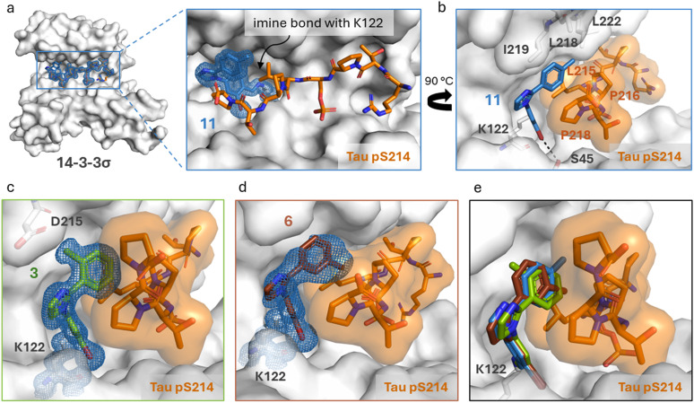
Protein-protein interactions (PPIs) are key regulators of various cellular processes. Modulating PPIs with small molecules has gained increasing attention in drug discovery, particularly targeting the 14-3-3 protein family, which interacts with several hundred client proteins and plays a central role in cellular networks. However, targeting a specific PPI of the hub protein 14-3-3, with its plethora of potential client proteins, poses a significant selectivity challenge. This not only involves the selectivity of 14-3-3 PPIs with other client proteins, but also the selective stabilization of a specific 14-3-3 binding site within a protein partner featuring several binding sites. The interaction of 14-3-3 with Tau, characterized by different phospho-site driven binding modes, forms a valuable, disease-relevant, 14-3-3 multivalent model PPI to explore this selectivity issue. This work presents the identification and early-stage optimization of small molecule fragment-like stabilizers for a specific binding site of the 14-3-3/Tau PPI. Using different biophysical assays, the stabilizing potency of the imine-bond forming molecules was mapped and X-ray crystallography studies provided structural data on the binding mode of the ternary complexes. Exploiting the unique topologies and functionalities of the different binding sites enabled the engineering of selectivity for this initial molecular glue matter for the pS214 binding site, over a second 14-3-3 binding site in Tau (pS324). These reversible covalent tool compounds will allow for the further exploration of the role of 14-3-3 in Tau aggregation.


Conflict of interest statement

The authors declare the following competing financial interest(s): L. B. and C. O. are scientific co-founders of Ambagon Therapeutics.

Figures

Fig. 1
Fig. 1. a Primary structure and domains of Tau protein, as well as the sequence of the bivalent Tau pS214–pS324 (pS2) peptide. b Schematic visualization of the different 14-3-3/Tau binding modes depending on the phosphorylation state of Tau with the corresponding binding affinity and aggregation propensity with a focus on the phosphorylation sites S214 and S324. Tau phosphorylated at only S214 or S324 leads analogously as the non-phosphorylated Tau to the formation of a 2 : 2 complex (14-3-3 monomer : Tau) instead of a 1 : 2 complex in case of bivalent Tau. The complex in which either both S214 and S324 are phosphorylated as well as the complex in which only S214 is phosphorylated is expected to be aggregation-resistant, while the other complexes have been reported to induce aggregation. Figure adapted from Chen et al. c Schematic visualization of a phosphosite-selective molecular glue to stabilize the 14-3-3/Tau pS214 interaction and potential to convey aggregation-resistance. d General chemotype used in the screening campaign to identify stabilizers of the 14-3-3/Tau pS2 peptide interaction and chemical structures of hit compound 1 and 2.
Fig. 2
Fig. 2. a (Left) Crystal structure of ternary complex 14-3-3σΔC/Tau pS214 peptide/1. (Middle) Zoom-in of the crystallized ternary complex 1 (blue sticks)/14-3-3σΔC (white surface)/Tau pS214 peptide (orange sticks) which shows the imine bond formation between 1 and K122 of 14-3-3σΔC. 2Fo–Fc electron density maps are contoured at 1σ. (Right) Zoom-in demonstrating the halogen-bond of 1 (blue sticks) with its bromo substituent to the hydroxy group of S45 residue of 14-3-3σΔC. The phenyl ring of 1 is positioned in a hydrophobic pocket shaped by residues L215, P216, and P218 of the Tau pS214 peptide (orange sticks) and L218 and I219 of 14-3-3σ (white sticks). Stacking of P218 on the interface of the phenyl group allows for the lone pair–π interaction between P218 and the π-electron system of phenyl ring. Potential chemical space created by 14-3-3 surrounding compound 1 is shown in white sticks. PDB ID: 9FS4. b Synthesis design of 2-substituted 1-arylimidazoles and synthesis route of compound library. General chemical structures of the three compound series 1–3 are shown.
Fig. 3
Fig. 3. a Dose–response fluorescence anisotropy data of compound titrations to 10 nM FITC-labeled Tau pS2 peptide and 100 nM 14-3-3ζ. Background (without 14-3-3ζ) subtracted, overnight measurement, mean and SD of technical duplicate (see ESI Fig. S5 for another replicate). b Bar plot representation of CC50 values of the compound titrations shown in (a), mean and SD of scientific duplicate. c Dose–response fluorescence anisotropy data of 14-3-3ζ titrations to 10 nM FITC-labeled Tau pS2 peptide and 250 μM compound, mean and SD of technical duplicate (see ESI Fig. S6 for another replicate). d Radar plot of stabilization factors at 250 μM compound 18 (SF250) from 14-3-3ζ FA protein titrations with 10 nM (Tau pS2 peptide) or 100 nM (13 other peptides) FITC-labeled peptide. Mean of three individually performed experiments (see ESI Fig. S8 for the protein titrations). e Differential scanning fluorimetry assay results. Identified melting temperatures (Tm) of 14-3-3γ alone (2.5 μM) and in the presence of Tau pS2 peptide (25 μM) and additionally with selected compounds (75 μM). Mean of three individually performed experiments. f 19F-NMR spectra (565 MHz, 298 K, 10 vol% D2O in H2O) of compound 21 (125 μM) at varying concentrations of 14-3-3γ (0–250 μM) indicates ligand binding to 14-3-3γ.
Fig. 4
Fig. 4. Crystal structures of methylated compounds in complex with 14-3-3σΔC/Tau pS214. a Front view of the ternary complex 14-3-3σΔC (white surface), Tau pS214 (orange sticks) and 11 (blue sticks). Zoom in of the crystallized ternary complex 11 (blue sticks)/14-3-3σΔC (white surface)/Tau pS214 (orange sticks) which shows the imine bond formation between 11 and K122 of 14-3-3σΔC. 2Fo–Fc electron density maps are contoured at 1σ. PDB ID: 9FVP. b Side view of the ternary complex 14-3-3σΔC (white surface), Tau pS214 (orange sticks) and 11 (blue sticks). c Side view of the ternary complex 14-3-3σΔC (white surface), Tau pS214 (orange sticks) and 3 (green sticks). 2Fo–Fc electron density maps are contoured at 1σ. PDB ID: 9FVH. d Side view of the ternary complex 14-3-3σΔC (white surface), Tau pS214 (orange sticks) and 6 (brown sticks). 2Fo–Fc electron density maps are contoured at 1σ. PDB ID: 9FVI. e Overlay of the three methylated compounds inside the 14-3-3σΔC/Tau pS214 complex.
Fig. 5
Fig. 5. a Phospho-site selective 14-3-3/Tau PPI stabilization. b SF250μM of selected compounds for stabilization of the 14-3-3γ complex with Tau pS214 and Tau pS324 peptides based on the internal DMSO reference. Mean of two separately performed experiments (see ESI Fig. S9 for the corresponding binding curves). c Differential scanning fluorimetry assay results. Identified melting temperatures (Tm) of 14-3-3γ in the presence of the Tau pS214 or pS324 peptide and compounds 1 and 18. Mean of two separately performed experiments. d and e 14-3-3γ titrations in the presence of increasing concentrations of compounds 1 (d) and 18 (e), in the presence of either 100 nM fluorescein-labeled Tau pS214 or fluorescein-labeled Tau pS324. Overnight measurement, mean and SD of scientific duplicate. f, SF250μM of the 14-3-3γ/Tau pS214 interaction plotted against compound concentrations. The data are derived from 2D titrations (d and e). The saturation of the ratio represents the α-factor. (h) Ratio of KD/apparent KD of the 14-3-3γ/Tau pS324 interaction plotted against compound concentrations. The data are derived from 2D titrations (figures d and e). The saturation of the ratio represents the α-factor.
Fig. 6
Fig. 6. a Chemical structure of compound 18. b Ternary structure of 14-3-3σΔC (white surface), Tau pS214 (orange sticks) and 18 (light orange sticks) complex. Zoom-in of the crystallized ternary complex 18 (light orange sticks)/14-3-3σΔC (white surface)/Tau pS214 (orange sticks) which shows the imine bond formation between 18 and K122 of 14-3-3σΔC and the occupation of the hydrophobic groove of 14-3-3 (white sticks). 2Fo–Fc electron density map (blue mesh) is contoured at 1σ (PDB ID: 9FVG). c Side view of an overlay of the ternary structure of 14-3-3σΔC (white surface)/Tau pS214 (orange stick)/18 (light orange sticks) complex with the binary 14-3-3σΔC/Tau pS214 (grey sticks) complex (PDB ID: 4FL5). The grey arrow represents the conformational change of the P218 residue due to compound 18.

References

    1. Stumpf M. P. H. Thorne T. De Silva E. Stewart R. Hyeong J. A. Lappe M. Wiuf C. Proc. Natl. Acad. Sci. U. S. A. 2008;105:6959–6964. doi: 10.1073/pnas.0708078105. - DOI - PMC - PubMed
    1. Koh G. C. K. W. Porras P. Aranda B. Hermjakob H. Orchard S. E. J. Proteome Res. 2012;11:2014–2031. doi: 10.1021/pr201211w. - DOI - PubMed
    1. Lu H. Zhou Q. He J. Jiang Z. Peng C. Tong R. Shi J. Signal Transduction Targeted Ther. 2020;5:213. doi: 10.1038/s41392-020-00315-3. - DOI - PMC - PubMed
    1. Nevola L. Giralt E. Chem. Commun. 2015;51:3302. doi: 10.1039/C4CC08565E. - DOI - PubMed
    1. Wells J. A. Mcclendon C. L. Nature. 2007;450:1001–1009. doi: 10.1038/nature06526. - DOI - PubMed

LinkOut - more resources