. 2025 Mar 7:18:1115-1129.
doi: 10.2147/JPR.S506569. eCollection 2025.

Electroacupuncture Inhibits NLRP3-Mediated Microglial Pyroptosis to Ameliorate Chronic Neuropathic Pain in Rats

Affiliations

Electroacupuncture Inhibits NLRP3-Mediated Microglial Pyroptosis to Ameliorate Chronic Neuropathic Pain in Rats

Wenyun Kui et al. J Pain Res. .

Abstract

Background: Patients with neuropathic pain (NP), caused by injury or disease of the somatosensory nervous system, usually suffer from severe pain. Our previous studies revealed that electroacupuncture (EA) stimulation could effectively improve NP. However, the underlying mechanisms of EA have not been fully clarified. This study aimed to investigate the specific mechanisms of EA in alleviating NP, focusing on the pyroptosis.

Materials and methods: Chronic Constriction Injury (CCI) model was established on the male Sprague-Dawley rats. CCI rats were treated with EA at acupoints GV20 and ST36 or/with the NOD-like receptor protein 3 (NLRP3) antagonist MCC950. EA treatment was administered for successive 14 days 7 days after the CCI surgery. The mechanical withdrawal threshold (MWT) and paw withdrawal latency (PWL) were performed during the experiment. At the end of the experiment, spinal cord segments and serum of rats were collected, ELISA detected the expression of inflammatory factors, immunofluorescence detected the microglia and neuron cells with pyroptosis biomarkers, and Western blot detected the NLRP3 pathway.

Results: EA treatment significantly alleviated pain hypersensitivity by increasing the MWT and PWL. Moreover, EA reduced levels of pro-inflammatory cytokines IL-1β and TNF-α in the spinal tissue. Mechanistically, the pyroptosis-related proteins, including NLRP3, N-GSDMD, Cleaved Caspase-1, IL-18 as well as IL-1β were downregulated by EA, indicating that EA attenuated the pyroptosis phenotype in NP rats. In particular, EA reduced the co-expression of NLRP3, Caspase-1 and N-GSDMD in microglia rather than in neuronal or astrocytic cells within the spinal cord of CCI rats. Pharmacological inhibition of NLRP3 inflammasome by MCC950 alleviates CCI-induced pain hypersensitivity while blocking EA's effect on anti-pyroptosis in CCI rats.

Conclusion: These findings demonstrate the EA ameliorates the neuroinflammation and pyroptosis to relieve chronic NP by suppressing NLRP3 inflammasome activation in microglia. EA may serve as a viable treatment therapy for chronic NP.

Keywords: NLRP3 inflammasome; electroacupuncture; neuroinflammation; neuropathic pain; pyroptosis.


Conflict of interest statement

The authors report no conflicts of interest in this work.

Figures
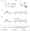
Figure 1
Figure 1
The establishment of the CCI induced NP modeling and EA’s intervention CCI rats. (A) Demonstration of the CCI surgery. (B) The intervention protocol of EA treatment. (C) The study protocol of EA treatment in CCI rats in experiment 1. (D) The study protocol of EA and MCC950 treatment in CCI rats in experiment 2.
Figure 2
Figure 2
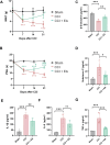
EA reduced pain hypersensitivity and suppressed the spinal inflammatory responses in CCI-induced NP rats. (A) Effects of EA on MWT induced by CCI surgery. (B) Effects of EA on PWL in CCI-induced NP models. *P < 0.05, ***P < 0.001. vs Sham group. #P < 0.05, ###P < 0.001 vs CCI group. n = 8. (C) The level of β-Endorphin in the spinal cord tissue of each group. (D) The level of SP in the serum of each group. (EG) The level of IL-1β, IL-6 and TNF-α in the spinal cord tissue of each group. *P < 0.05, **P < 0.01, ***P < 0.001. ns, no significant difference. n = 5.
Figure 3
Figure 3
EA attenuated CCI-induced pyroptosis phenotype in the spinal cord. (A) The protein expression of NLRP3, N-GSDMD, Cleaved Caspase-1, IL-18 and IL-1β in the spinal cord of each group. β-actin was used as an internal control. (B and F) The relative expression of NLRP3, N-GSDMD, Cleaved Caspase-1 and IL-18 protein in each group. Data presented as means ± s.d. *P < 0.05, **P < 0.01, ***P < 0.001. n = 3. (G and I) The mRNA expression levels of the pyroptosis-related gene of NLRP3, IL-18, IL-1β, respectively. Data presented as means ± s.d. *P < 0.05, **P < 0.01, ***P < 0.001. n = 3.
Figure 4
Figure 4
EA inhibited the pyroptosis in microglia following CCI operation. (A) Representative images of immunofluorescent analysis of Iba-1+ (green), NLRP3+ (red) maker, and DAPI (blue) staining of nuclei in the spinal cord dorsal horn among groups. Scale bars, 50 μm. (B) Representative images of immunofluorescent analysis of Iba-1+ (green), Caspase-1+ (red) maker, and DAPI (blue) staining of nuclei in the spinal cord dorsal horn among groups. Scale bars, 50 μm. (C) Representative images of immunofluorescent analysis of Iba-1+ (green), N-GSDMD+ (red) maker, and DAPI (blue) staining of nuclei in the spinal cord dorsal horn among groups. Scale bars, 50 μm. Quantification analysis the number of NLRP3/Iba-1+ (D), Caspase-1/Iba-1+ (E), N-GSDMD/Iba-1+ (F) in the spinal cord dorsal horn. Data presented as means ± s.d. *P < 0.05, **P < 0.01, ***P < 0.001, n = 3.
Figure 5
Figure 5
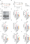
Inhibition of NLRP3 inflammasome activation relieved CCI‑induced pain behaviour and abolished the anti-pyroptosis effect of EA. (A and B) The effects of EA on MWT and PWL in CCI-induced NP models. n = 8. Data presented as means ± s.d. ***P < 0.001. vs Sham group. ##P < 0.01, ###P < 0.001. vs CCI group. (C) The protein expression of NLRP3, N-GSDMD, Cleaved Caspase-1, IL-18 and IL-1β in the spinal cord of each group. β-actin was used as an internal control. (D and H) The relative expression of NLRP3, N-GSDMD, Cleaved Caspase-1, IL-18 and IL-1β protein in each group. Data presented as means ± s.d. *P < 0.05, **P < 0.01, ***P < 0.001. n = 3. (I and K) The level of IL-6, IL-1β and TNF-α in the spinal cord of each group. Data presented as means ± s.d. *P < 0.05, ***P < 0.001. ns, no significant difference.n = 5.
Figure 6
Figure 6
A schematic diagram illustrating the mechanism of EA treatment in peripheral nerve injury-induced NP.

References

    1. Petzke F, Tölle T, Fitzcharles MA, Häuser W. Cannabis-Based Medicines and Medical Cannabis for Chronic Neuropathic Pain. CNS Drugs. 2022;36(1):31–44. doi:10.1007/s40263-021-00879-w - DOI - PMC - PubMed
    1. Bannister K, Sachau J, Baron R, Dickenson AH. Neuropathic Pain: mechanism-Based Therapeutics. Annu Rev Pharmacol Toxicol. 2020;60(1):257–274. doi:10.1146/annurev-pharmtox-010818-021524 - DOI - PubMed
    1. Wei YJ, Hsieh CF, Huang YT, Huang MS, Fang TJ. The influence of integrated geriatric outpatient clinics on the health care utilization of older people. BMC Geriatr. 2020;20(1):379. doi:10.1186/s12877-020-01782-7 - DOI - PMC - PubMed
    1. Gilron I, Baron R, Jensen T. Neuropathic pain: principles of diagnosis and treatment. Mayo Clin Proc. 2015;90(4):532–545. doi:10.1016/j.mayocp.2015.01.018 - DOI - PubMed
    1. Dai SP, Yang CC, Chin Y, Sun WH. T cell death-associated gene 8-mediated distinct signaling pathways modulate the early and late phases of neuropathic pain. iScience. 2024;27(10):110955. doi:10.1016/j.isci.2024.110955 - DOI - PMC - PubMed

LinkOut - more resources