. 2025 May 13;148(5):1652-1664.
doi: 10.1093/brain/awae374.

Parvalbumin neurons mediate neurological phenotypes of anti-NMDAR encephalitis

Affiliations

Parvalbumin neurons mediate neurological phenotypes of anti-NMDAR encephalitis

Yi-Fan Feng et al. Brain. .

Abstract

Patients with anti-N-methyl-D-aspartate receptor (anti-NMDAR) encephalitis, often present with severe psychiatric symptoms, yet the neuropathological mechanisms underlying their cognitive deficits remain insufficiently understood. In this study, we constructed an animal model using anti-NMDAR IgG purified from the serum of patients with anti-NMDAR encephalitis, and we used IgG obtained from healthy individuals as a control. Daily administration of anti-NMDAR IgG into the medial prefrontal cortex (mPFC) of mice for 7 days resulted in cognitive impairments resembling clinical symptoms, which spontaneously resolved 30 days after discontinuing the injections. Immunohistochemical staining and electrophysiological testing of parvalbumin neurons in the mPFC treated with anti-NMDAR IgG revealed significant cellular morphological damage, reduced excitability, synaptic dysfunction and a loss of NMDAR antagonist-induced gamma oscillations. Application of optogenetic and pharmacogenetic techniques to activate parvalbumin neurons in the mPFC successfully reversed the cognitive impairments observed in the anti-NMDAR-IgG-treated mice. Single-cell sequencing of anti-NMDAR-IgG-treated parvalbumin neurons identified differentially expressed genes and pathways related to synapses and neuronal development, offering potential targets for therapeutic intervention. Additionally, we showed that these alterations in parvalbumin neurons were not confined to the mPFC, as similar changes were detected in the hippocampus after anti-NMDAR IgG injections. In summary, our findings elucidate distinct alterations in parvalbumin neurons during the pathogenesis of anti-NMDAR encephalitis, providing preclinical rationale for exploring approaches to modulate parvalbumin neuronal function to treat anti-NMDAR encephalitis.

Keywords: NMDAR; cognitive impairments; gamma oscillations; medial prefrontal cortex; parvalbumin neurons; rescue.


Conflict of interest statement

The authors report no competing interests.

Figures
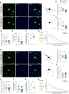
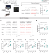
Figure 1
Figure 1
The morphology of mPFC parvalbumin neurons was reversible after anti-NMDAR IgG injection. (AG) After 7 days of antibody injection, the morphology, scale bar = 20 μm (A), number of primary branches (B), number of secondary branches (C), dendrite length (D), PV+ boutons intensity (E), PV+ neuron density (F) and Sholl analysis (G) of medial prefrontal cortex (mPFC) PV neurons. Scale bar = 20 μm, n = 18–25 neurons of 6–7 mice. For PV+ neuron density (F), n = 6 mice. (HN) Thirty days after antibody injection, the morphology, scale bar = 20 μm (H), number of primary branches (I), number of secondary branches (J), dendrite length (K), PV+ boutons intensity (L), PV+ neuron density (M) and Sholl analysis (N) of mPFC PV neurons. Scale bar = 20 μm, 1 pixel = 0.19 μm, n = 23–29 neurons of six mice. For PV+ neuron density (M), n = 6 mice. *P < 0.05, **P < 0.01, ***P < 0.001. ns = not significant; PV = parvalbumin.
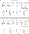
Figure 2
Figure 2
Anti-NMDAR IgG specifically reduced the excitability of mPFC parvalbumin neurons and attenuated their inhibitory effect on pyramidal neurons. (A) Schematic diagrams of patch-clamp recordings and evoked action potentials in pyramidal neurons of C57/B6J mice after 7 days of antibody injection. (B) Excitability of pyramidal neurons, n = 12 neurons from six mice. (C) Schematic diagrams of patch-clamp recordings and evoked action potentials in parvalbumin (PV) neurons of PV-Cre mice after 7 days of antibody injection. (D) Excitability of PV neurons, n = 12 neurons from six mice. **P < 0.01, ****P < 0.0001. (E) Representative traces of spontaneous excitatory postsynaptic currents (sEPSCs) of PV neurons, n = 16–19 neurons from six mice. (F) Cumulative probability plots of the inter-event interval of sEPSCs, analysed using Kruskal-Wallis one-way ANOVA. (G) Cumulative probability plots of the amplitude of sEPSCs, analysed using Kruskal-Wallis one-way ANOVA. (H) Average frequency of sEPSCs in mice injected with anti-NMDAR or control IgG. ****P < 0.0001. (I) Average amplitude of sEPSCs in mice injected with anti-NMDAR or control IgG. **P < 0.01. (J) Schematic diagram of paired pulse ratio (PPR) recordings in PV neurons after antibody injection. (K) Statistical analysis of PPR, n = 23–32 neurons from six mice. ****P < 0.0001.
Figure 3
Figure 3
Anti-NMDAR IgG abolished the NMDAR antagonist-induced elevation of low gamma oscillations. (A) Schematic diagram showing the timeline for MK-801 challenge and local field potential (LFP) electrode position. Scale bar = 200 μm. (B and C) Gamma baseline without MK-801 injection, n = 5. (DI) Comparison of oscillations in various frequency bands before and after intraperitoneal injection of MK-801. (D) Example LFP data of mice. (EI) Statistical analysis of normalized LFP power in the low gamma, high gamma, theta, alpha and beta bands. n = 5, *P < 0.05, **P < 0.01.
Figure 4
Figure 4
Activation of mPFC parvalbumin neurons reversed cognitive impairment in mice injected with anti-NMDAR IgG. (A) Schematic diagram showing the timeline for optogenetic behavioural experiments. (B and C) Optogenetic activation of the medial prefrontal cortex (mPFC) PV neurons on Y-maze and novel object recognition (NOR) tests in mice after 7 days of antibody injection. n = 7, *P < 0.05. (D and E) Optogenetic activation of the mPFC PV on Y-maze and NOR tests in mice 30 days after antibody injection. n = 7. (F) Schematic diagram showing the timeline for chemogenetic behavioural experiments. (G and H) Chemogenetic activation of the mPFC PV neurons on Y-maze and NOR tests in mice after 7 days of antibody injection. n = 6, *P < 0.05, **P < 0.01. (I and J) Chemogenetic activation of the mPFC PV neurons on Y-maze and NOR tests in mice 30 days after antibody injection. n = 5–6.
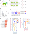
Figure 5
Figure 5
Single-cell sequencing after injection of control IgG/anti-NMDAR IgG into the medial prefrontal cortex. (A and B) A uniform manifold approximation and projection (UMAP) displaying cells from the medial prefrontal cortex (mPFC), categorized into 11 cell types using recognized markers and highly enriched genes. (C) Identification of major neurons in the mPFC. Pvalb = parvalbumin (PV) neurons; SST = somatostatin neurons; VIP = vasoactive intestinal peptide neurons. (D) Volcano map of differentially expressed genes between the anti-NMDAR IgG group and control IgG group. (E) Differentially expressed signalling pathways in each cluster. (F) Top signalling pathway enriched in Gene Ontology (GO) analysis. (G and H) Cell communication between clusters when PV neurons act as ligands (G) and receptors (H), n = 3.

References

    1. Hughes EG, Peng X, Gleichman AJ, et al. . Cellular and synaptic mechanisms of anti-NMDA receptor encephalitis. J Neurosci. 2010;30:5866–5875. - PMC - PubMed
    1. Finke C, Kopp UA, Prüss H, Dalmau J, Wandinger KP, Ploner CJ. Cognitive deficits following anti-NMDA receptor encephalitis. J Neurol Neurosurg Psychiatry. 2012;83:195–198. - PMC - PubMed
    1. Gibson LL, McKeever A, Coutinho E, Finke C, Pollak TA. Cognitive impact of neuronal antibodies: Encephalitis and beyond. Transl Psychiatry. 2020;10:304. - PMC - PubMed
    1. Dalmau J, Armangué T, Planagumà J, et al. . An update on anti-NMDA receptor encephalitis for neurologists and psychiatrists: Mechanisms and models. Lancet Neurol. 2019;18:1045–1057. - PubMed
    1. Planaguma J, Leypoldt F, Mannara F, et al. . Human N-methyl D-aspartate receptor antibodies alter memory and behaviour in mice. Brain. 2015;138(Pt 1):94–109. - PMC - PubMed