

Modeling of transendothelial transport
Abstract
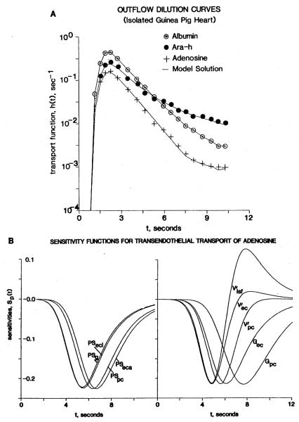
Capillary-tissue exchange of inert hydrophilic solutes in the heart occurs through aqueous channels, the clefts between endothelial cells (ECs). For adenosine (and other vasoactive agents and substrates), there is also transport across the plasmalemma of the ECs. The multiple-indicator dilution technique comparing tracer adenosine flux with that of 9-beta-D-arabinofuranosylhypoxanthine (an analog that is not transported by the nucleoside carrier) can be used to estimate the conductance of the facilitated transport mechanism, which is equivalent to a permeability-surface area product. Analysis by using a model of exchanges among capillary, EC, interstitium, and myocardial cells suggests that the abluminal surface of the ECs is also highly permeable to adenosine. The inference is that ECs may be an important component of a system for adenosine exchange and regulation in the heart.
Figures
References
-
- Bassingthwaighte JB, Chaloupka M, Wang CY. Transport by endothelial cells in vivo: model analysis from indicator dilution after single transcapillary passage. Federation Proc. 1983;42:580. (abstr.)
-
- Bassingthwaighte JB, Goresky CA. Modeling in the analysis of solute and water exchange in the microvasculature. In: Renkin EM, Michel CC, editors. Handbook of physiology, Section 2, The cardiovascular system, Vol. IV, Microcirculation, Part 1. Am. Physiol. Soc.; Bethesda: 1984. pp. 549–626.
-
- Bassingthwaighte JB, Knopp TJ, Hazelrig JB. A concurrent flow model for capillary-tissue exchanges. In: Crone C, Lassen NA, editors. Capillary permeability. Munksgaard; Copenhagen: 1970. pp. 60–80.
-
- Bassingthwaighte JB, Lenhoff AM, Stephenson JL. A slidingelement algorithm for rapid solution of spatially distributed convection-permeation models. Biophys. J. 1984;45:175a.
Publication types
MeSH terms
Substances
Grants and funding
LinkOut - more resources
Full Text Sources